©The Author(s) 2020.
World J Clin Cases. Nov 6, 2020; 8(21): 5446-5456
Published online Nov 6, 2020. doi: 10.12998/wjcc.v8.i21.5446
Published online Nov 6, 2020. doi: 10.12998/wjcc.v8.i21.5446
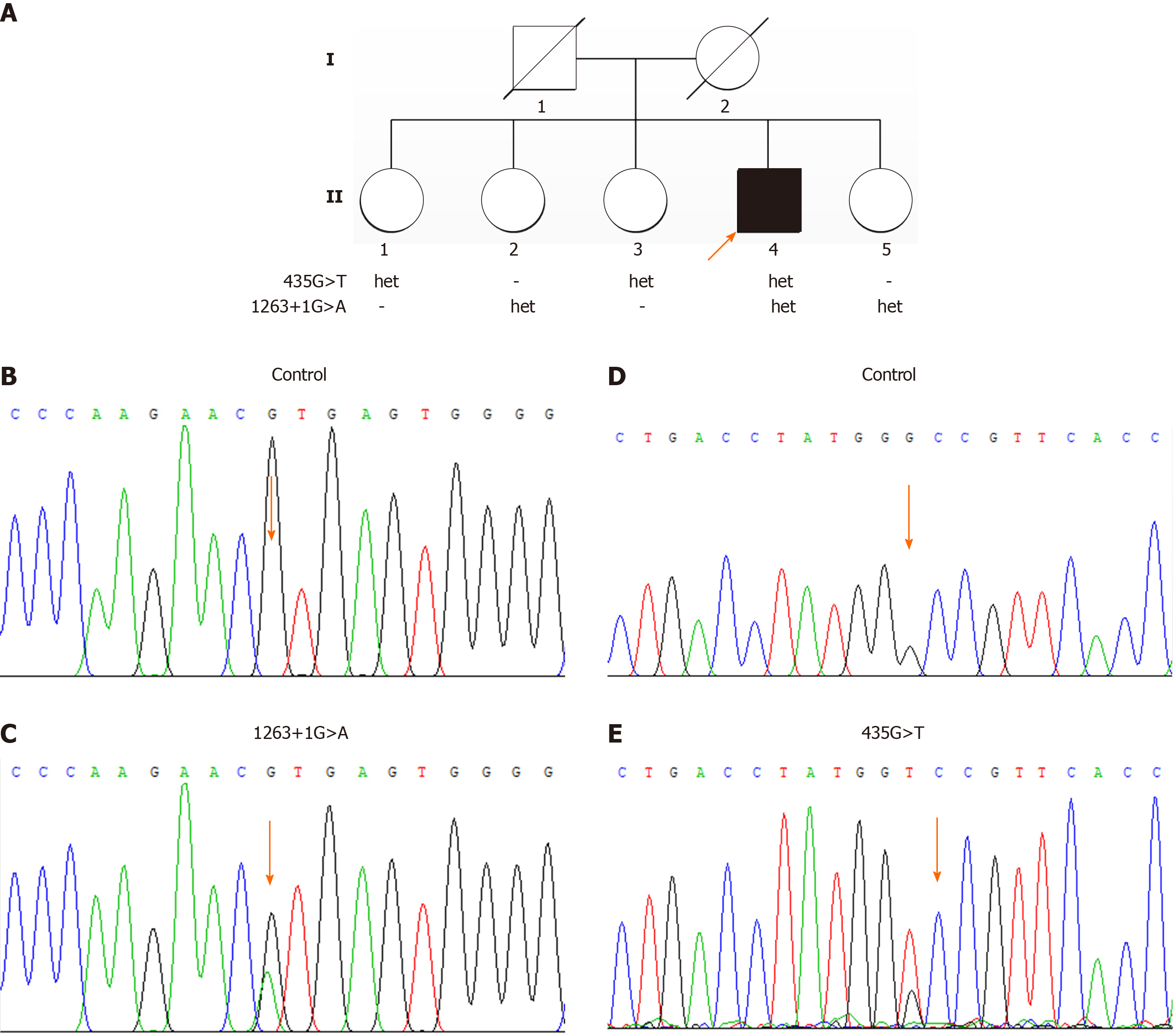
Figure 1 The pedigree of the family and the genetic test results.
A: The pedigree of the family with compound heterozygous CYP27A1 mutations; B and D: The normal control sequences; C: The variant in intron 7 (c.1263+1G>A); E: The variant in exon 2 (c.435G>T, p.Gly145Gly). Family members II:2 and II:5 were carriers of c.1263+1G>A and family members II:1 and II:3 were the carriers of c.435G>T. Family member II:4 carried the compound heterozygous CYP27A1 mutations.
Figure 2 Clinical and imaging data of the patient.
A: The dark skin and poor mental state of the patient; B: Arrowheads show the nodules on the bilateral tibial tubercles; C: Arrowheads show the enlarged Achilles tendons; D: B-ultrasound of the urinary system, showing the hypoechoic area (arrowhead) in the bladder which can move with body position; E: X-ray of the lower limbs showing swelling of the soft tissue below the bilateral tibial tuberosities (arrowheads); F: Sagittal T1-weighted magnetic resonance (MR) image showing fusiform swelling of the right Achilles tendon and upper medial malleolus flexor tendon, abnormal thickening of the plantar fat, and a small amount of exudation around the fascia in front of the Achilles tendon; G: Axial T2-weighted MR image suggesting mild ventricular enlargement and cerebral atrophy; H: Axial fluid-attenuated inversion recovery MR image showing an abnormal hyperintense signal around the ventricle posterior horn bilaterally (arrowheads).
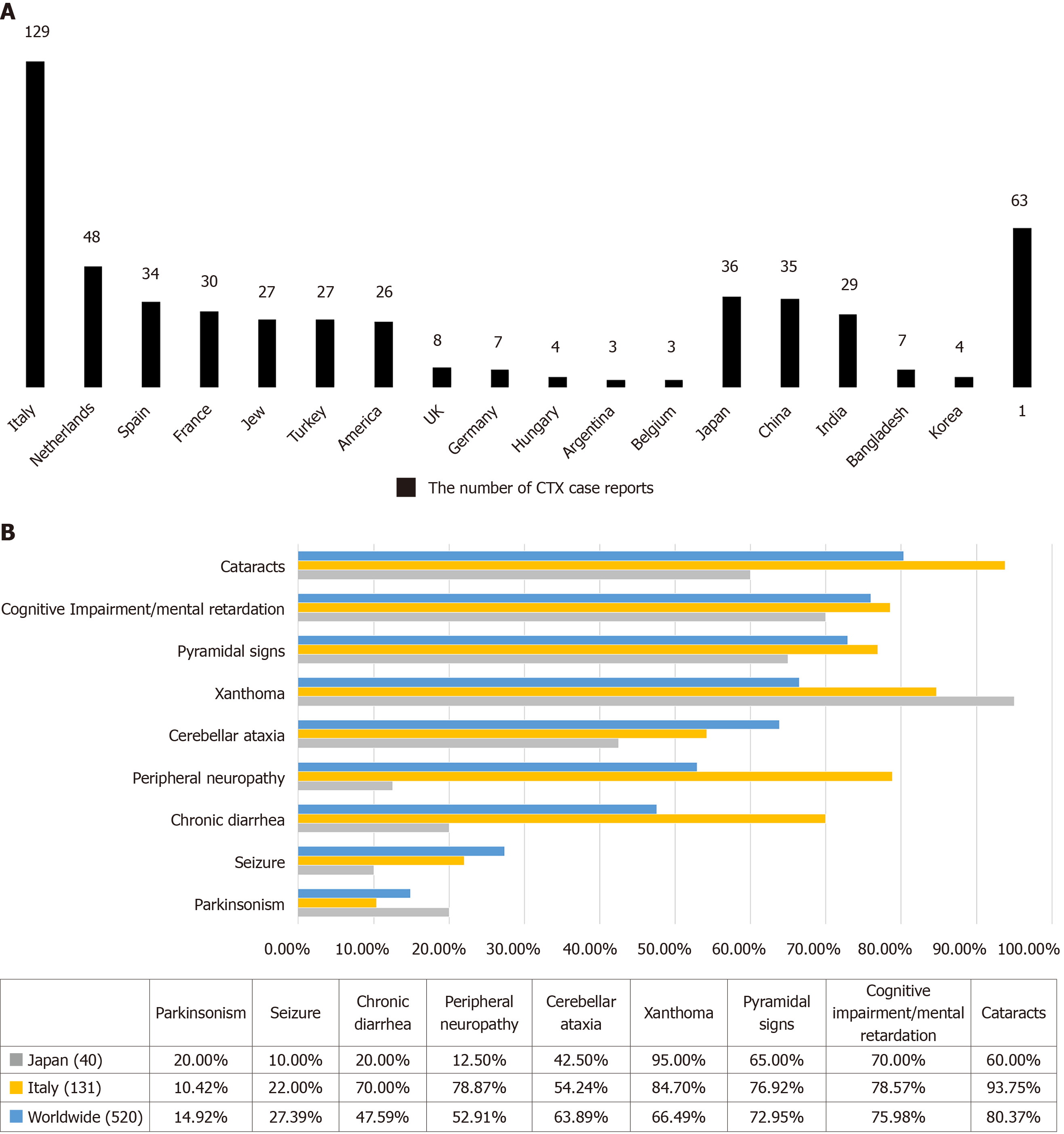
Figure 3 Main symptoms in cerebrotendinous xanthomatosis case reports from different ethnic groups.
1Refers to other origins and unclassified cases. A: The number of cerebrotendinous xanthomatosis (CTX) case reports from different origins in the past 30 years; B: Comparison of the frequencies of the main symptoms in CTX patients from all over the world.
- Citation: Cao LX, Yang M, Liu Y, Long WY, Zhao GH. Chinese patient with cerebrotendinous xanthomatosis confirmed by genetic testing: A case report and literature review . World J Clin Cases 2020; 8(21): 5446-5456
- URL: https://www.wjgnet.com/2307-8960/full/v8/i21/5446.htm
- DOI: https://dx.doi.org/10.12998/wjcc.v8.i21.5446


