©The Author(s) 2020.
World J Clin Cases. Nov 6, 2020; 8(21): 5334-5340
Published online Nov 6, 2020. doi: 10.12998/wjcc.v8.i21.5334
Published online Nov 6, 2020. doi: 10.12998/wjcc.v8.i21.5334
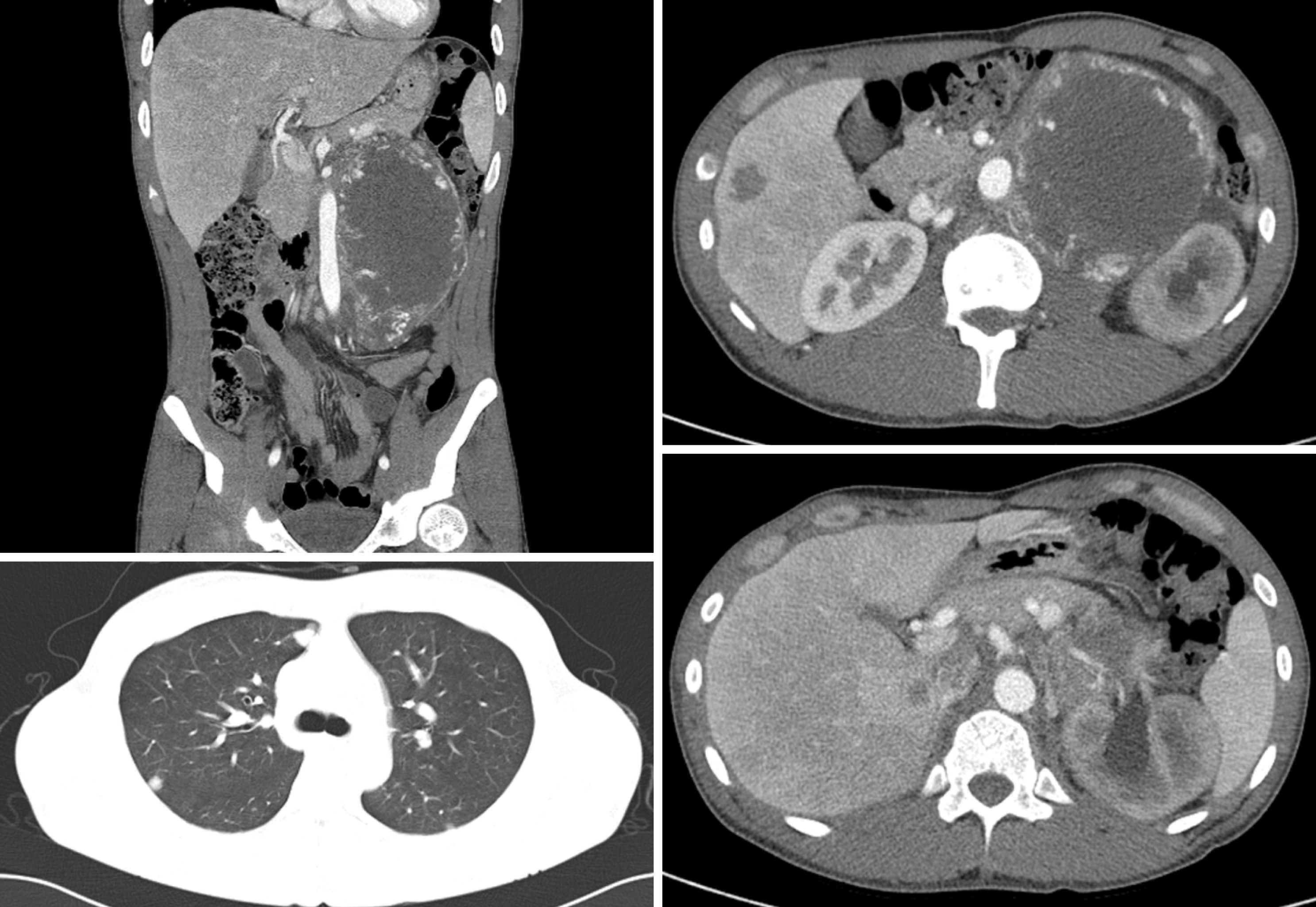
Figure 1 A baseline computed tomography scan showing a huge left retroperitoneal mass and multiple metastatic lesions on the liver and lung.
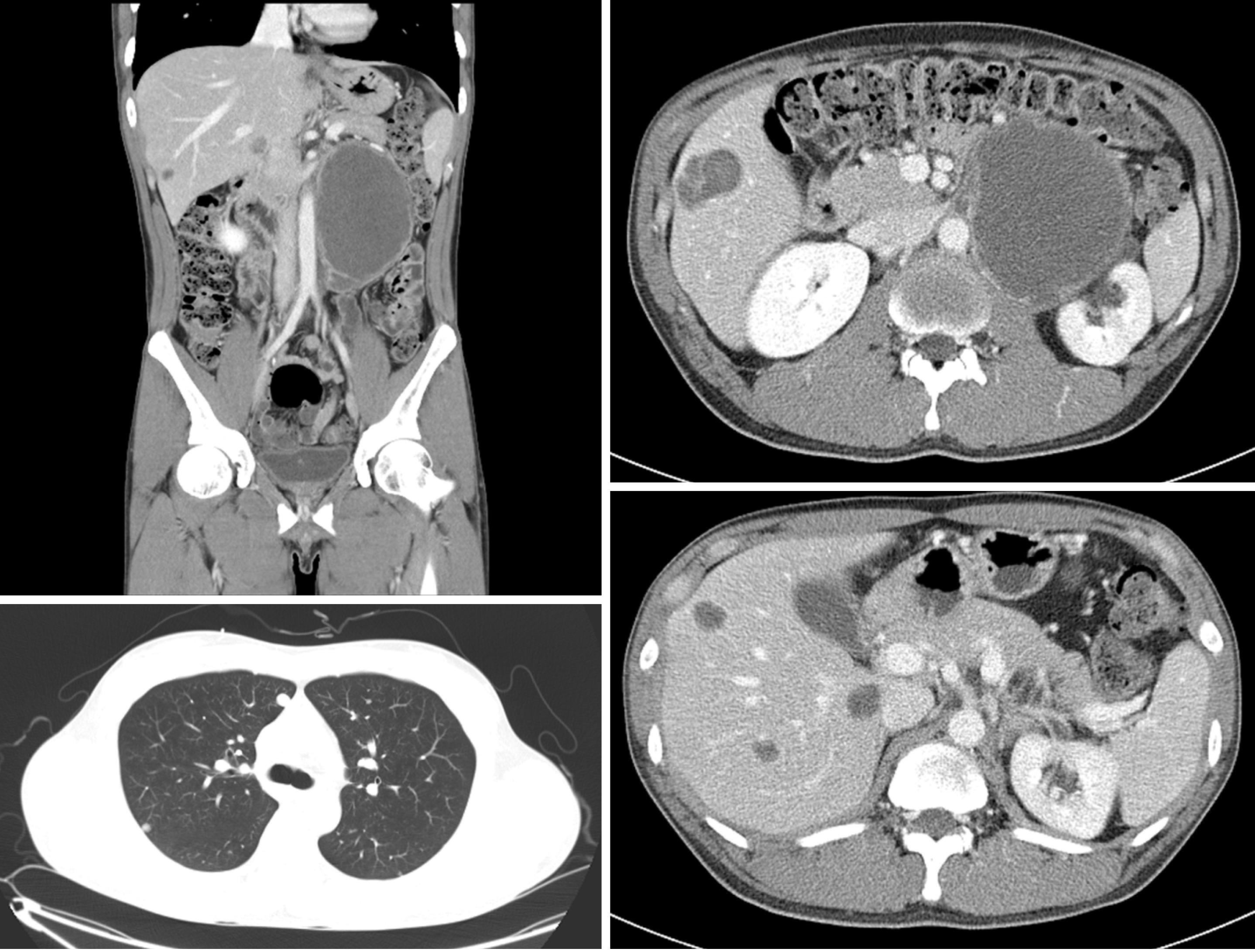
Figure 2 A follow-up computed tomography scan after 2 cycles of bleomycin, etoposide, and cisplatin chemotherapy, showing that the left retroperitoneal mass had decreased in size, but that liver metastatic lesions had increased in size and a new lesion in S6 had developed.
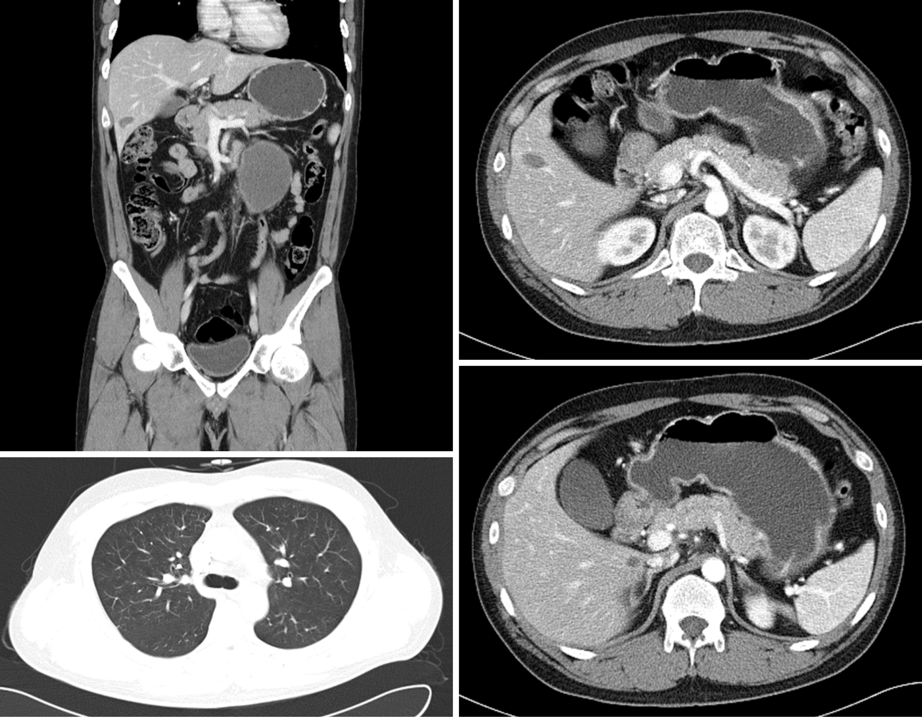
Figure 3 A follow-up computed tomography scan, after 8 cycles of etoposide, methotrexate, actinomycin D, cyclophosphamide, and vincristine regimen, showing that the left retroperitoneal mass and metastatic liver lesions had markedly decreased in size and that metastatic lung lesions had nearly disappeared.
Figure 4 Macroscopic features of the resected specimen, showing that tumors removed from liver and retroperitoneum, were relatively well-marginated and round-shaped lesions.
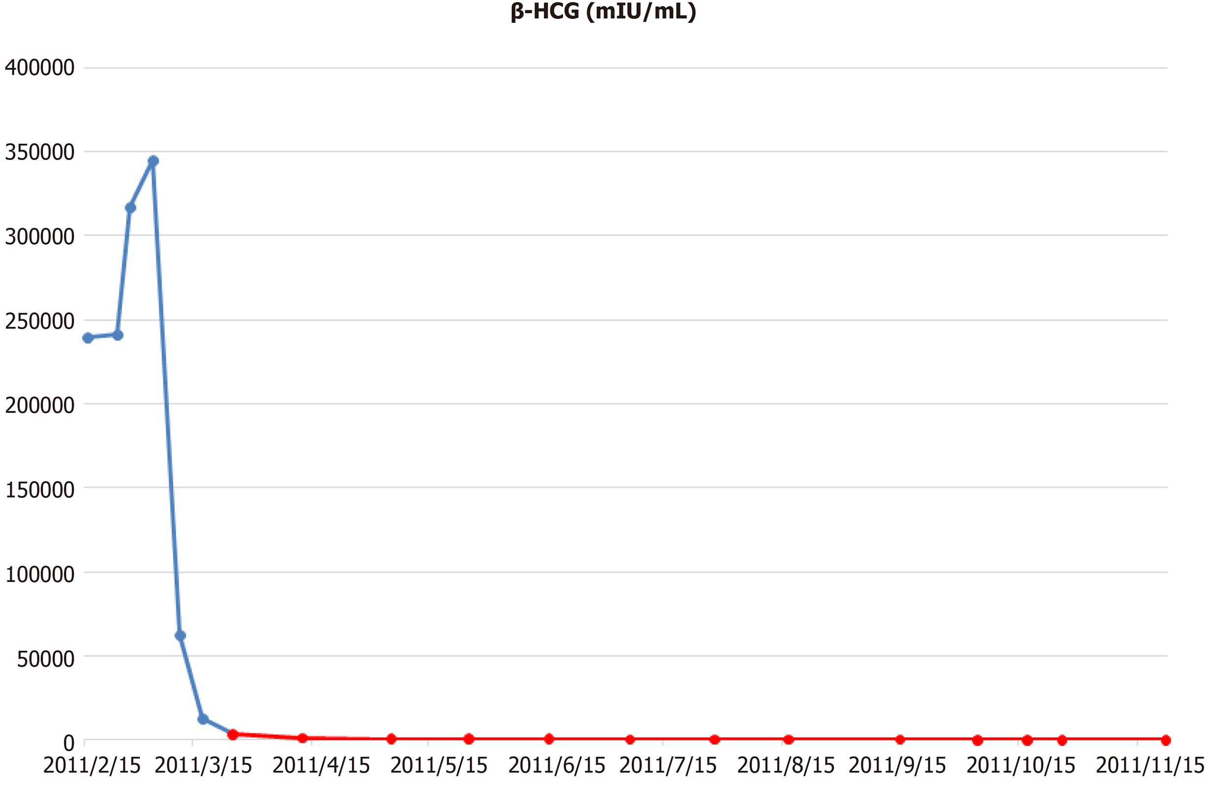
β-HCG: β human chorionic gonadotropin.
- Citation: Yun J, Lee SW, Lim SH, Kim SH, Kim CK, Park SK. Successful treatment of a high-risk nonseminomatous germ cell tumor using etoposide, methotrexate, actinomycin D, cyclophosphamide, and vincristine: A case report. World J Clin Cases 2020; 8(21): 5334-5340
- URL: https://www.wjgnet.com/2307-8960/full/v8/i21/5334.htm
- DOI: https://dx.doi.org/10.12998/wjcc.v8.i21.5334


