©The Author(s) 2020.
World J Clin Cases. Nov 6, 2020; 8(21): 5296-5303
Published online Nov 6, 2020. doi: 10.12998/wjcc.v8.i21.5296
Published online Nov 6, 2020. doi: 10.12998/wjcc.v8.i21.5296
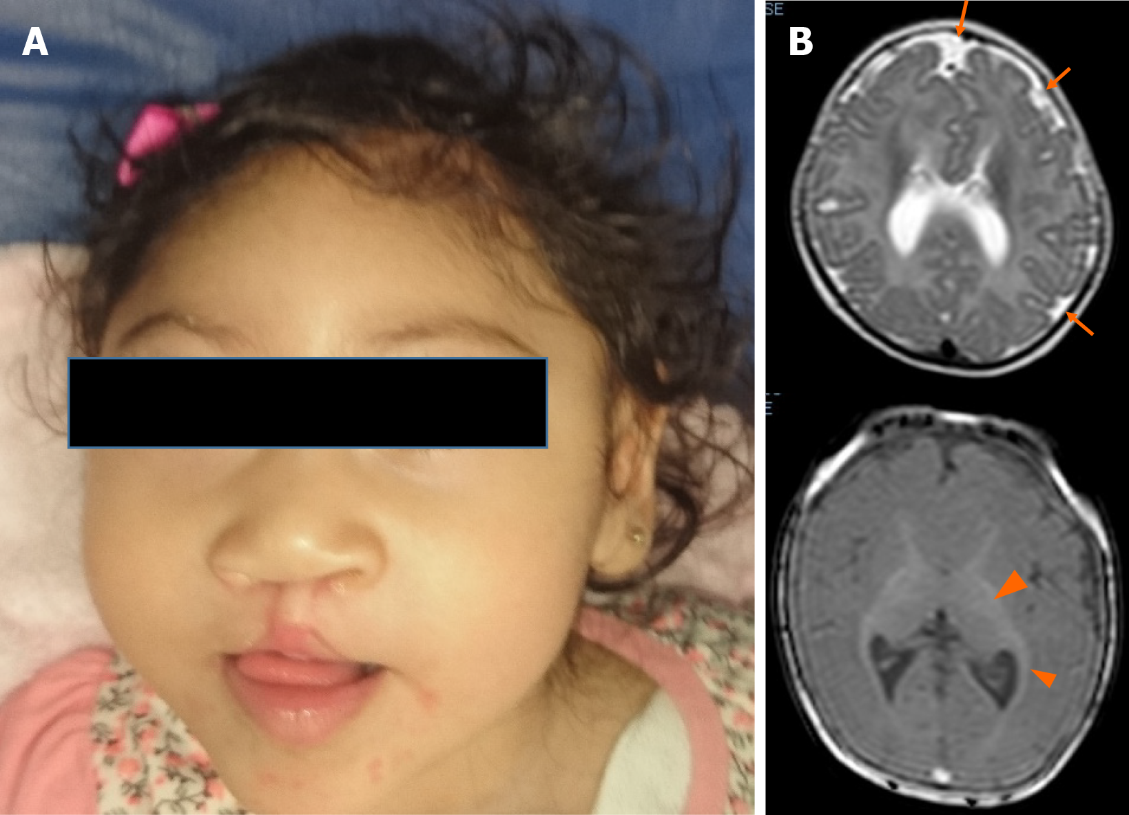
Figure 1 Patient at 1-year-old.
A: Microcephaly, upward-slanting palpebral fissures, depressed nasal bridge, bulbous nose, bilateral cleft lip, and palate are showed; B: The brain magnetic resonance scan (at five months) shows cortical atrophy, simplified gyral cortical patterns (orange arrow) and band heterotopia (arrow ahead).
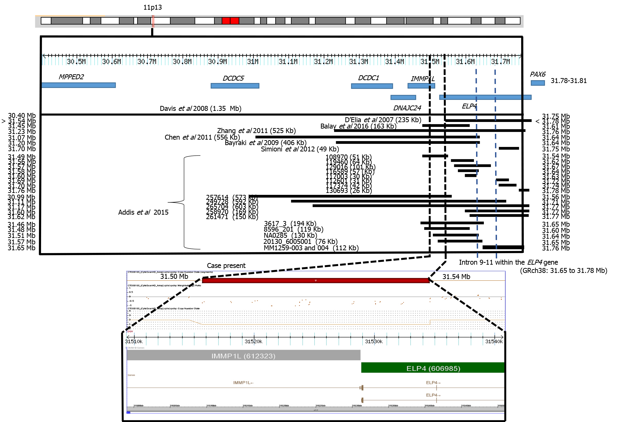
Figure 2 CytoScan high definition array and schematic representation of the result.
The figure shows chromosome 11p13 and the relative positions of the MPPED2, DCDC5, DCDC1, DNAJC24, IMMP1L, ELP4, and PAX6 genes within the deleted interval. A partial molecular karyotype of the submicroscopic on chromosome 11 detected with the CytoScan high definition array is also illustrated. A single copy of the 40 Kb region was identified on log2 ratio analysis. Some affected patients with deletions in the 11p13 region are also shown.
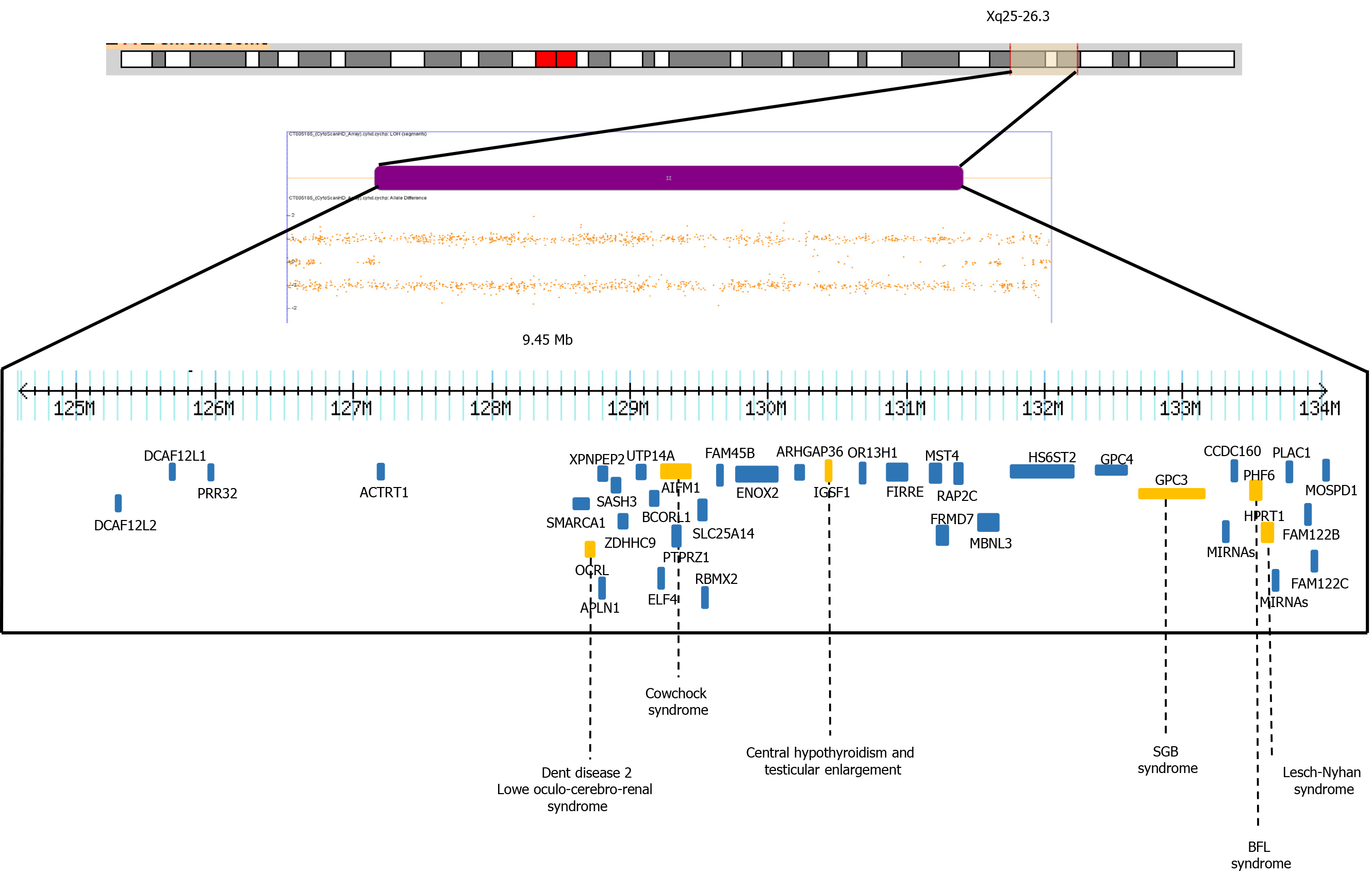
Figure 3 Depiction of allele peaks on chromosome Xq25-q26 shows homozygous (top and bottom bands) and heterozygous (middle band) allele peak bands.
Note the loss of the middle band showing loss of heterozygosity of Xq25-26.3. The genes related to X-linked diseases (OCRL, AIFM1, IGSF1, GPC3, PHF6, HPRT1) (orange) such as Dent disease 2, Lowe Oculo-Cerebro-Renal syndrome, cowchock syndrome, central hypothyroidism and testicular enlargement, Simpson-Golabi-Behmel syndrome, Borjeson-Forssman-Lehman syndrome and Lesch-Nyhan are shown.
- Citation: Toral-Lopez J, González Huerta LM, Messina-Baas O, Cuevas-Covarrubias SA. Submicroscopic 11p13 deletion including the elongator acetyltransferase complex subunit 4 gene in a girl with language failure, intellectual disability and congenital malformations: A case report . World J Clin Cases 2020; 8(21): 5296-5303
- URL: https://www.wjgnet.com/2307-8960/full/v8/i21/5296.htm
- DOI: https://dx.doi.org/10.12998/wjcc.v8.i21.5296


