©The Author(s) 2020.
World J Clin Cases. Nov 6, 2020; 8(21): 5188-5202
Published online Nov 6, 2020. doi: 10.12998/wjcc.v8.i21.5188
Published online Nov 6, 2020. doi: 10.12998/wjcc.v8.i21.5188
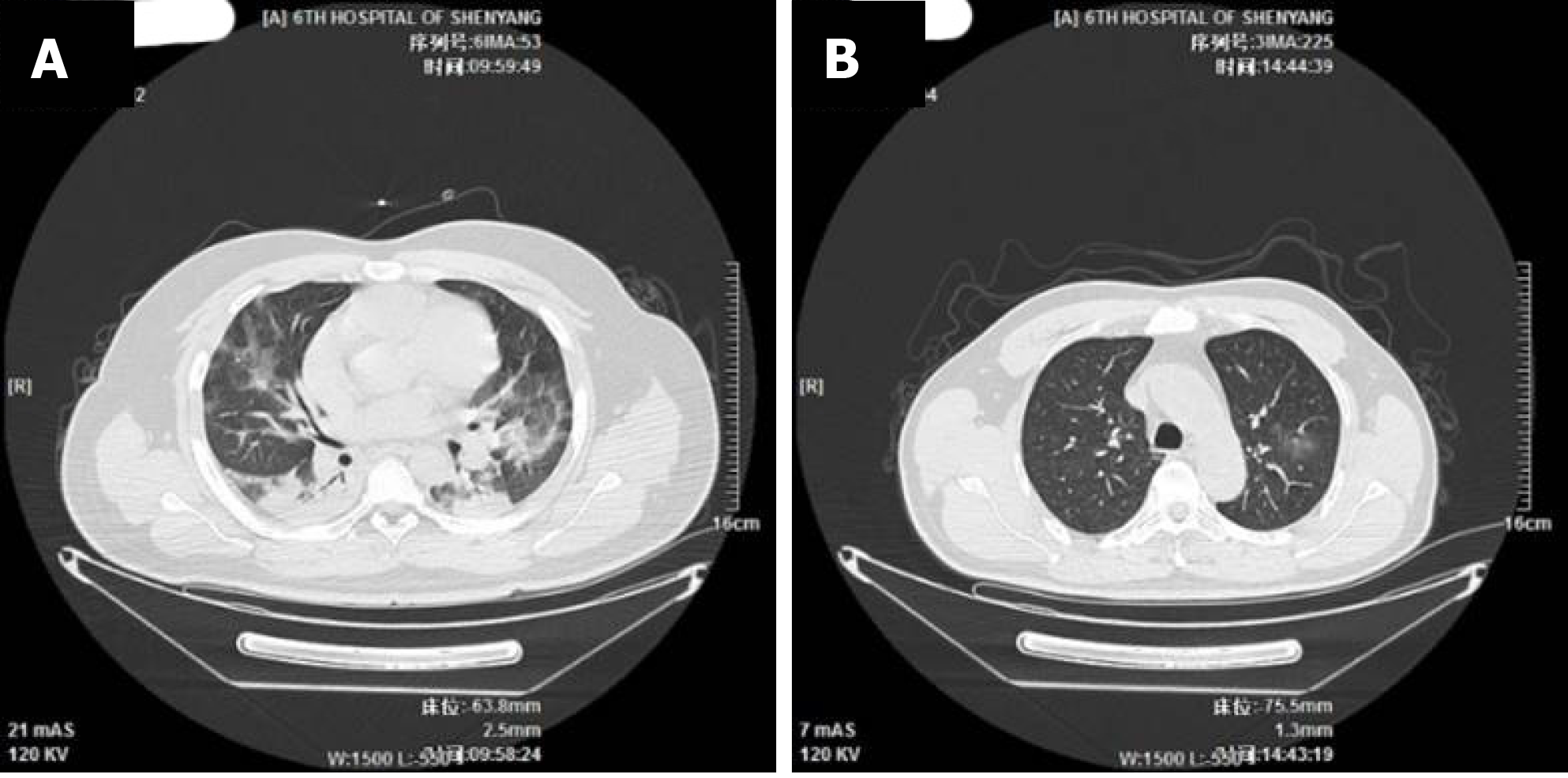
Figure 1 Comparison of lung computed tomography images of a serious case and a mild case.
A: Multiple indistinct patchy opacities in both lungs; large areas of ground-glass opacity and consolidative opacity; large areas of consolidative opacity in the basal segments of the lower lobes of both lungs, with air bronchogram visible inside. Lesions are serious in the middle and outer zones [26-year-old male patient, computed tomography (CT) taken on February 2, 2020, the 7th d after disease onset]; B: A few indistinct patchy opacities in the upper lobe of the left lung, middle lobe and lower lobe of the right lung (47-year-old male patient, CT taken on February 4, 2020, the 5th d after disease onset).
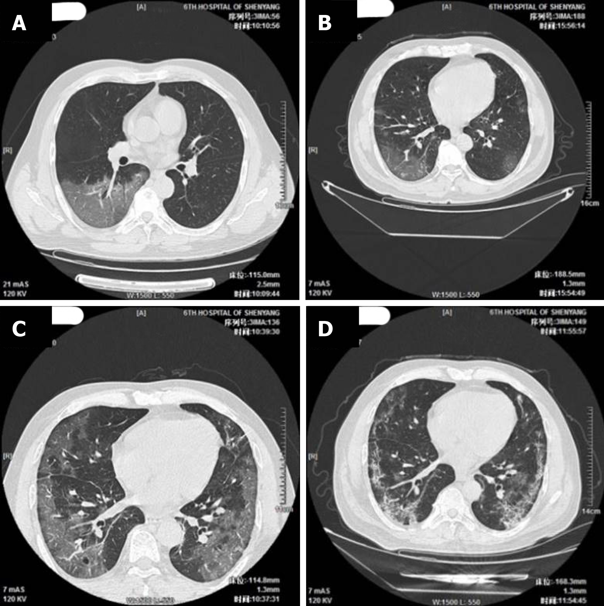
Figure 2 Progression of pneumonia as observed in the lung computed tomography images of a serious case (51-year-old male patient).
A: Multiple small patchy opacities and large areas of ground-glass opacity in both lungs, particularly in the outer zones and under the pulmonary pleurae; larger area of lesion in the basal segment of the lower right lobe, with air bronchogram and vascular thickening visible inside [computed tomography (CT) taken on February 3, 2020, the 7th d after disease onset]; B: Multiple small patchy opacities and large areas of ground-glass opacity in both lungs, particularly in the middle and outer zones of bilateral lung fields and under the pulmonary pleurae; the lesions have slightly increased in number and size (CT taken on February 5, 2020, the 9th d after disease onset); C: Multiple small patchy opacities and large areas of ground-glass opacity in both lungs, particularly in the middle and outer zones and under the pulmonary pleurae; new patchy opacities and large areas of ground-glass opacity can be observed in the bilateral lower lobes and the right middle lobe (CT taken on February 10, 2020, the 14th d after disease onset); D: Multiple small patchy opacities and large areas of ground-glass opacity in both lungs, particularly in the middle and outer zones and under the pulmonary pleurae; the scope of the lesions shows a clear reduction (CT taken on February 17, 2020, the 21st d after disease onset).
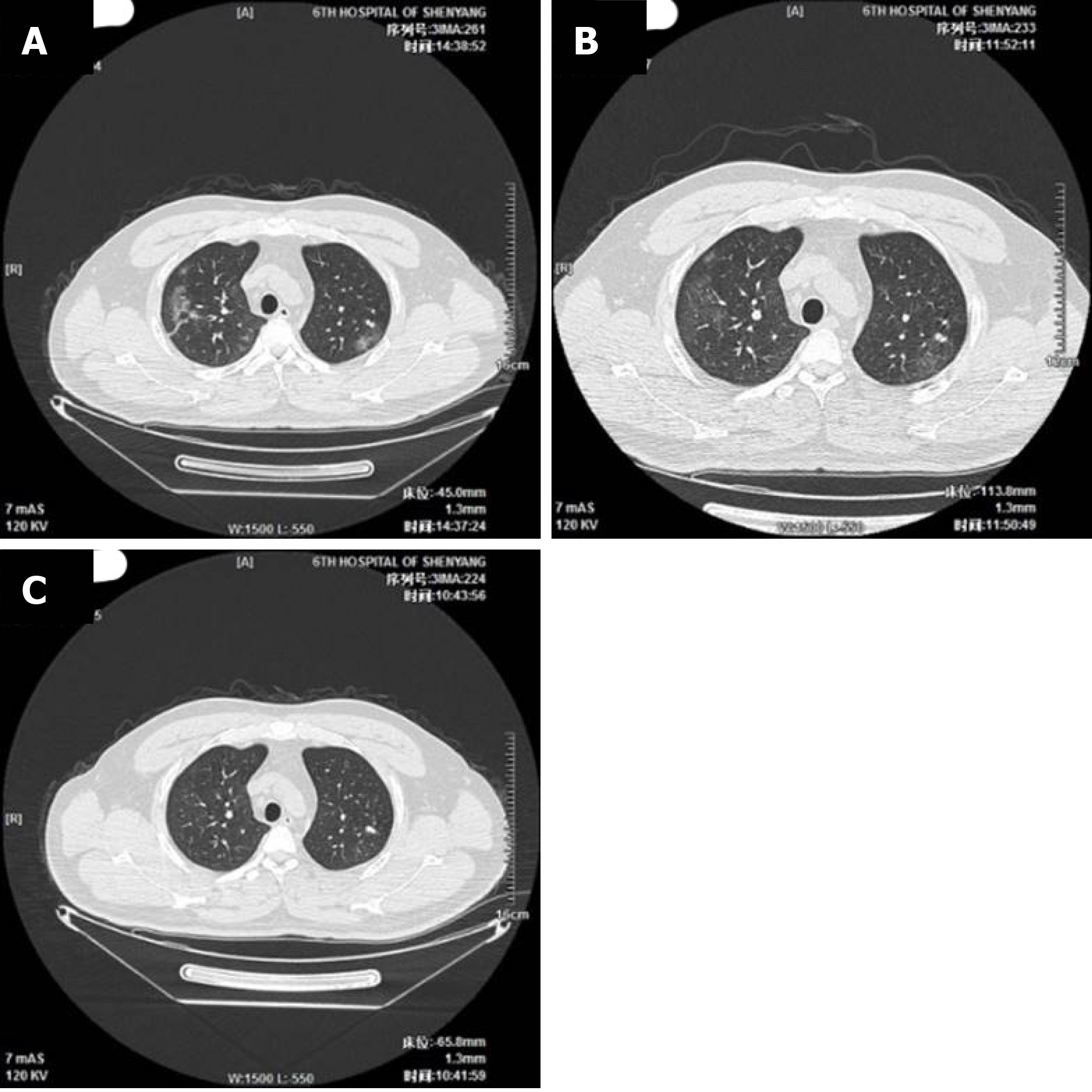
Figure 3 Development of pneumonia as observed on the lung computed tomography images of a mild case (39-year-old male patient).
A: Multiple indistinct patchy opacities, ground-glass opacities, and stripe-like opacities in both lungs, particularly in the right lung [computed tomography (CT) taken on February 4, 2020, the 8th d after disease onset]; B: Multiple indistinct patchy opacities, ground-glass opacities, and stripe-like opacities in both lungs, particularly in the right lung; the lesions show a reduction in size (CT taken on February 7, 2020, the 11th d after disease onset); C: A few light patchy opacities are scattered in the middle and outer zones of the right upper lobe and bilateral lower lobes; the lesions show a clear reduction in size (CT taken on February 15, 2020, the 19th d after disease onset).
- Citation: Wang JB, Wang HT, Wang LS, Li LP, Xv J, Xv C, Li XH, Wu YH, Liu HY, Li BJ, Yu H, Tian X, Zhang ZY, Wang Y, Zhao R, Liu JY, Wang W, Gu Y. Epidemiological and clinical characteristics of fifty-six cases of COVID-19 in Liaoning Province, China. World J Clin Cases 2020; 8(21): 5188-5202
- URL: https://www.wjgnet.com/2307-8960/full/v8/i21/5188.htm
- DOI: https://dx.doi.org/10.12998/wjcc.v8.i21.5188


