Copyright
©The Author(s) 2020.
World J Clin Cases. Oct 26, 2020; 8(20): 5062-5069
Published online Oct 26, 2020. doi: 10.12998/wjcc.v8.i20.5062
Published online Oct 26, 2020. doi: 10.12998/wjcc.v8.i20.5062
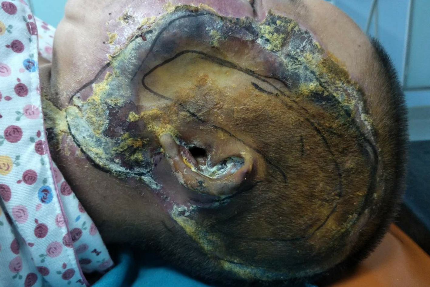
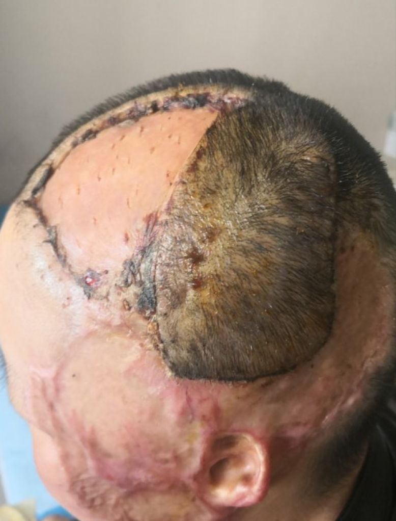
Figure 1 Left temporal and facial burn wounds form black scabs.
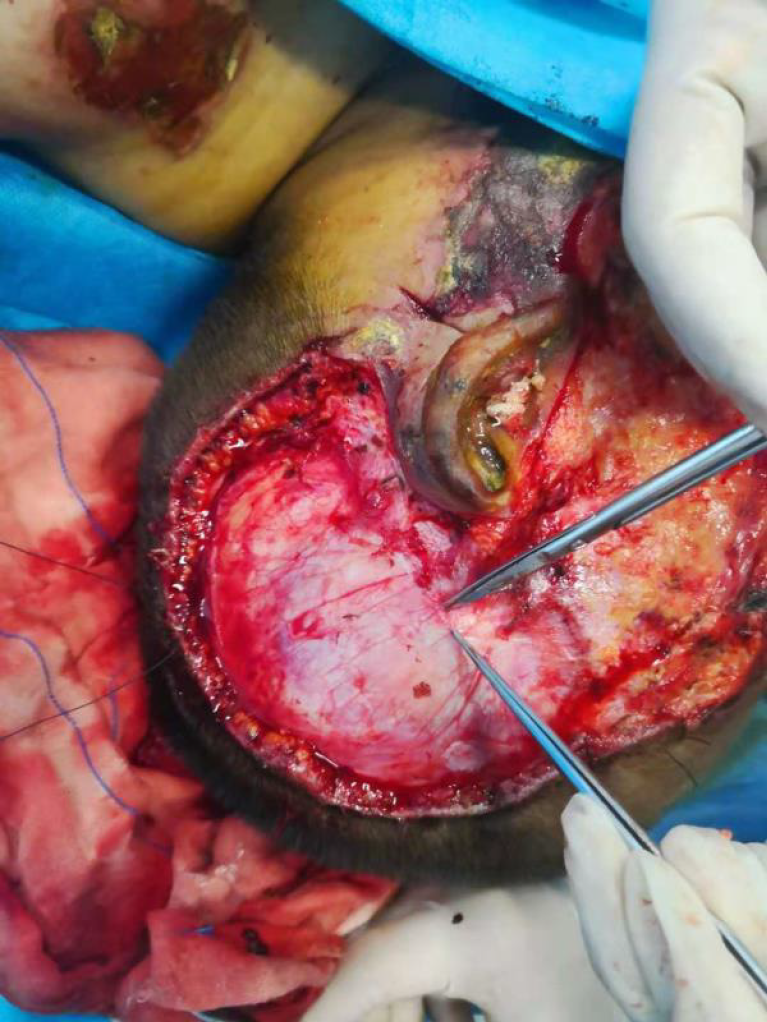
Figure 2 During the procedure, the left area of the skin tissue was removed, and the papillary muscle is seen.
The left facial skin tissue was removed to the SMARS fascia.
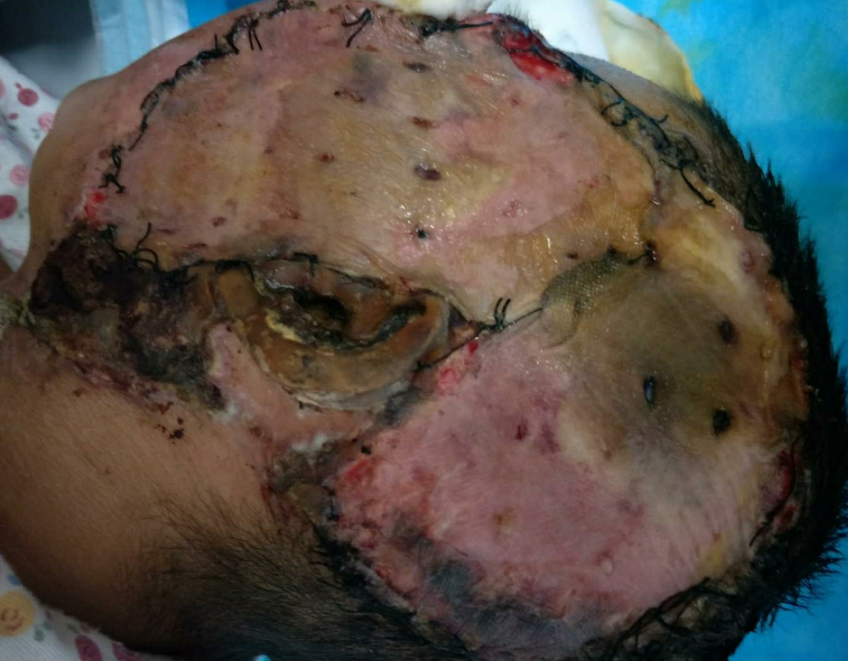
Figure 3 The left area of the skin (approximately 3 cm × 3 cm) showed poor survival.
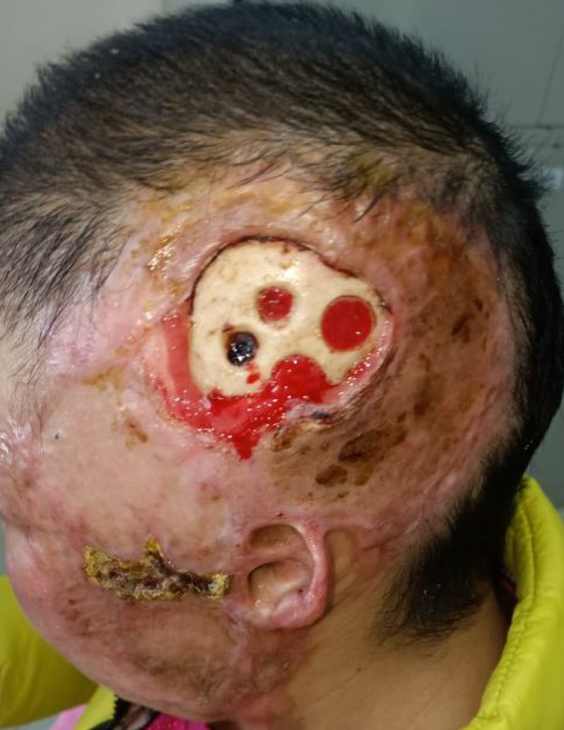
Figure 4 Granulation tissue seen after drilling of the left temporal skull.
Figure 5 Rotating flap covering the exposed bone.
- Citation: Shen CM, Li Y, Liu Z, Qi YZ. Effective administration of cranial drilling therapy in the treatment of fourth degree temporal, facial and upper limb burns at high altitude: A case report. World J Clin Cases 2020; 8(20): 5062-5069
- URL: https://www.wjgnet.com/2307-8960/full/v8/i20/5062.htm
- DOI: https://dx.doi.org/10.12998/wjcc.v8.i20.5062


