Copyright
©The Author(s) 2020.
World J Clin Cases. Oct 26, 2020; 8(20): 5013-5018
Published online Oct 26, 2020. doi: 10.12998/wjcc.v8.i20.5013
Published online Oct 26, 2020. doi: 10.12998/wjcc.v8.i20.5013
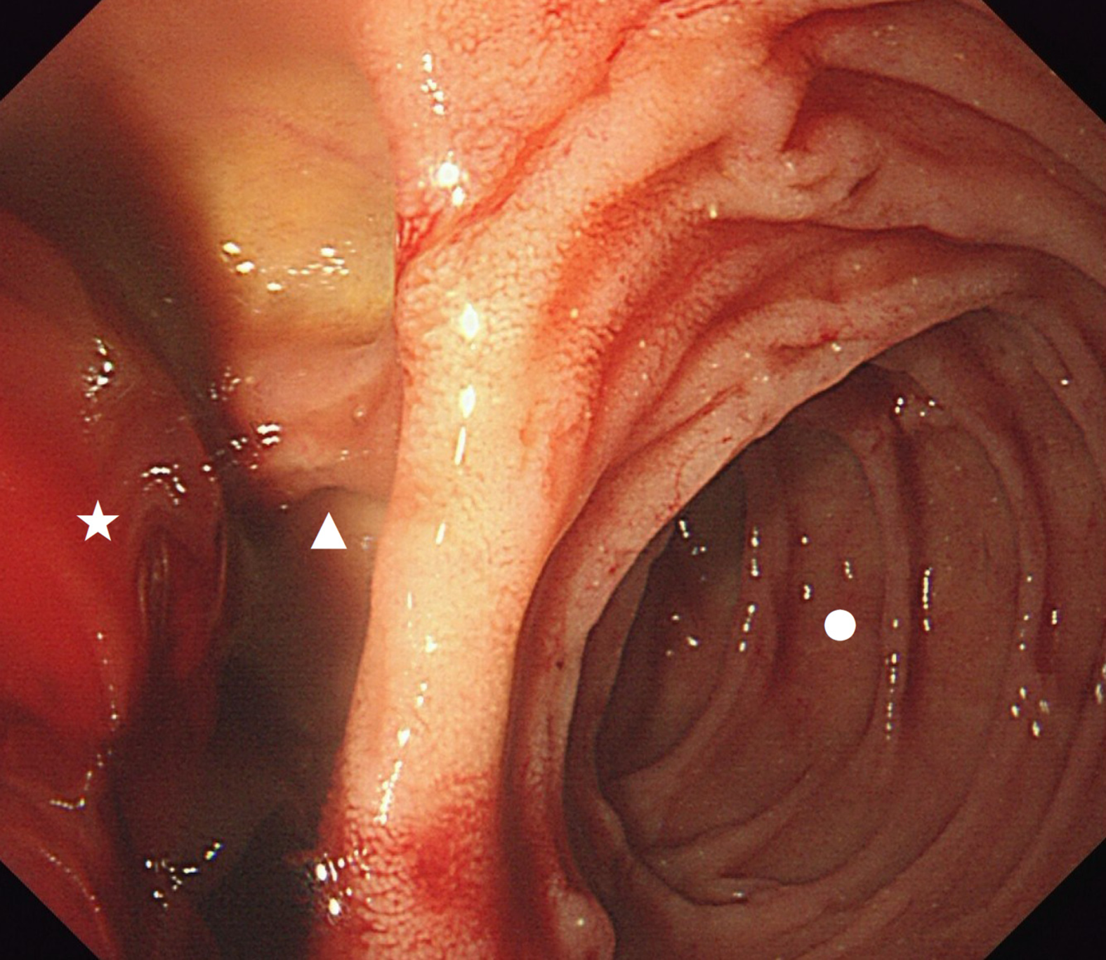
Figure 1 Endoscopic view of the diverticulum located in the descending duodenum.
Circle: Descending duodenum; Triangle: Diverticulum; Pentagram: Blood clot.
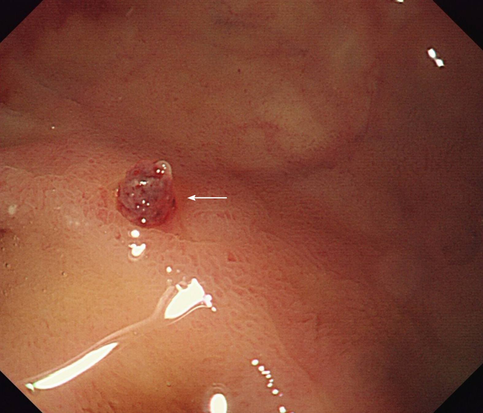
Figure 2 Endoscopic view of the isolated protruding artery of Dieulafoy disease after washing away the blood clot.
Arrow: Artery stump.
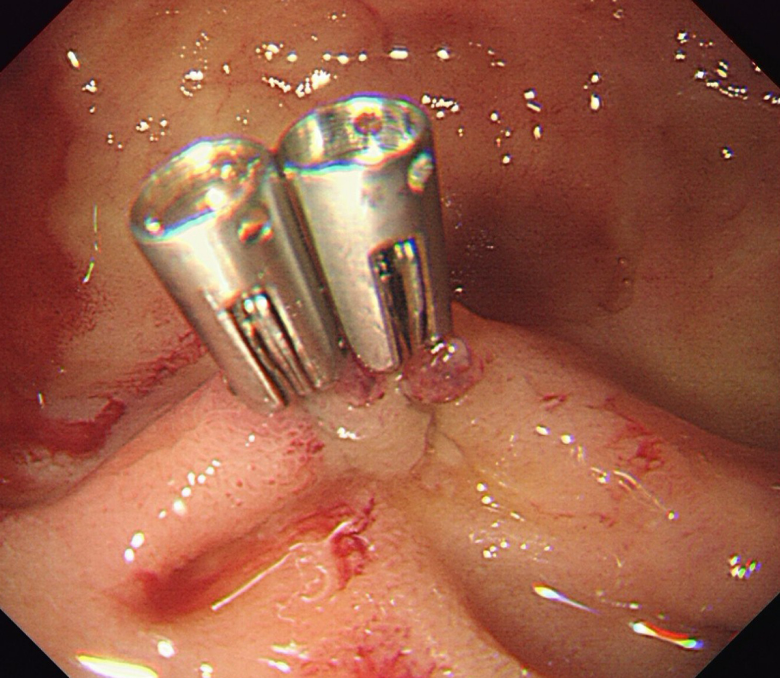
Figure 3 Two titanium clips were inserted to clamp the vessel stump.
- Citation: He ZW, Zhong L, Xu H, Shi H, Wang YM, Liu XC. Massive gastrointestinal bleeding caused by a Dieulafoy’s lesion in a duodenal diverticulum: A case report. World J Clin Cases 2020; 8(20): 5013-5018
- URL: https://www.wjgnet.com/2307-8960/full/v8/i20/5013.htm
- DOI: https://dx.doi.org/10.12998/wjcc.v8.i20.5013


