Copyright
©The Author(s) 2020.
World J Clin Cases. Oct 26, 2020; 8(20): 4981-4985
Published online Oct 26, 2020. doi: 10.12998/wjcc.v8.i20.4981
Published online Oct 26, 2020. doi: 10.12998/wjcc.v8.i20.4981
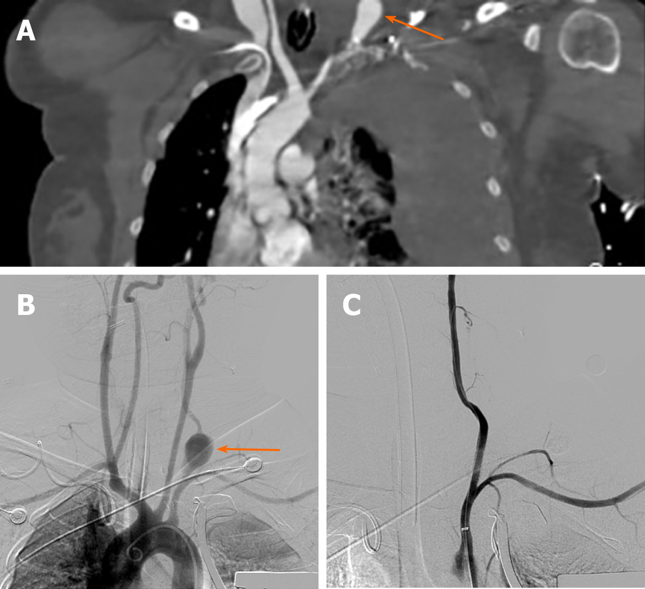
Figure 1 Coronal reconstruction of the contrast-enhanced chest CT (A) and digital subtraction angiography of the vertebral artery and endovascular stent implantation (B and C).
A: Pseudoaneurysm formation in the initial segment of the left vertebral artery (arrow) and a large amount of effusion in the left thoracic cavity; B: Contrast medium overflow at the start of the left vertebral artery (arrow), suggesting the formation of a pseudoaneurysm; C: The signs of vertebral arterial contrast medium spillage disappeared after stent implantation.
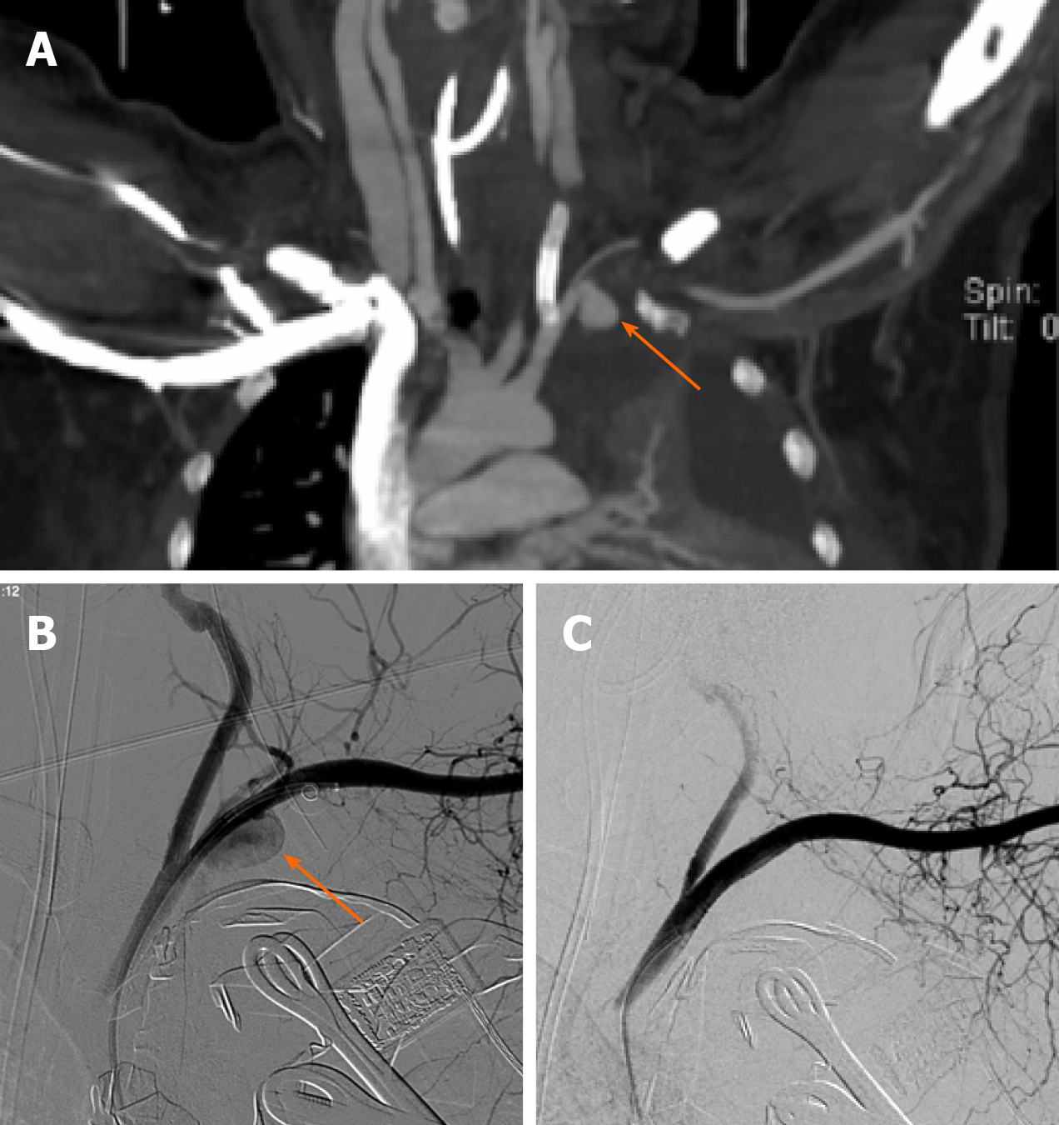
Figure 2 Coronal reconstruction of the contrast-enhanced chest CT (A) and digital subtraction angiography of the left subclavian artery and stent implantation.
A: Pseudoaneurysm formation at the start of the left subclavian artery (arrow) and endovascular stent implantation in the left vertebral artery. B: Contrast medium overflow at the start of the left subclavian artery, suggesting the formation of a pseudoaneurysm (arrow); C: After the stent graft was implanted, the contrast medium spillage disappeared.
- Citation: Huang W, Zhang GQ, Wu JJ, Li B, Han SG, Chao M, Jin K. Catastrophic vertebral artery and subclavian artery pseudoaneurysms caused by a fishbone: A case report. World J Clin Cases 2020; 8(20): 4981-4985
- URL: https://www.wjgnet.com/2307-8960/full/v8/i20/4981.htm
- DOI: https://dx.doi.org/10.12998/wjcc.v8.i20.4981


