Copyright
©The Author(s) 2020.
World J Clin Cases. Oct 26, 2020; 8(20): 4930-4937
Published online Oct 26, 2020. doi: 10.12998/wjcc.v8.i20.4930
Published online Oct 26, 2020. doi: 10.12998/wjcc.v8.i20.4930
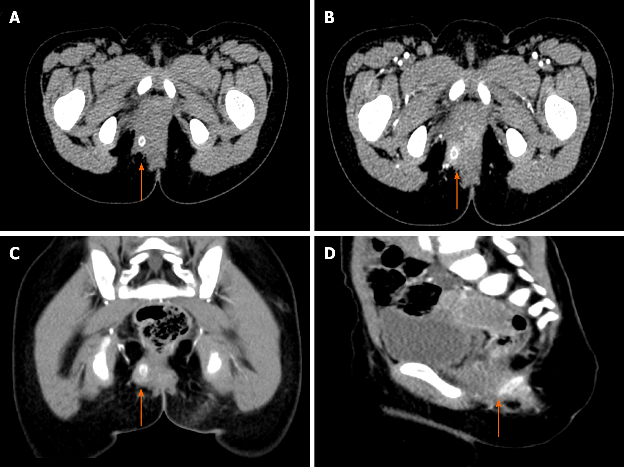
Figure 1 Preoperative abdominal computed tomography scan with intravenous contrast media administration of patient 1.
Axial view (A) and intravenous contrast-enhanced axial (B), coronal (C) and sagittal (D) views showing a foreign body (arrowheads) with uneven density and brim enhancement.
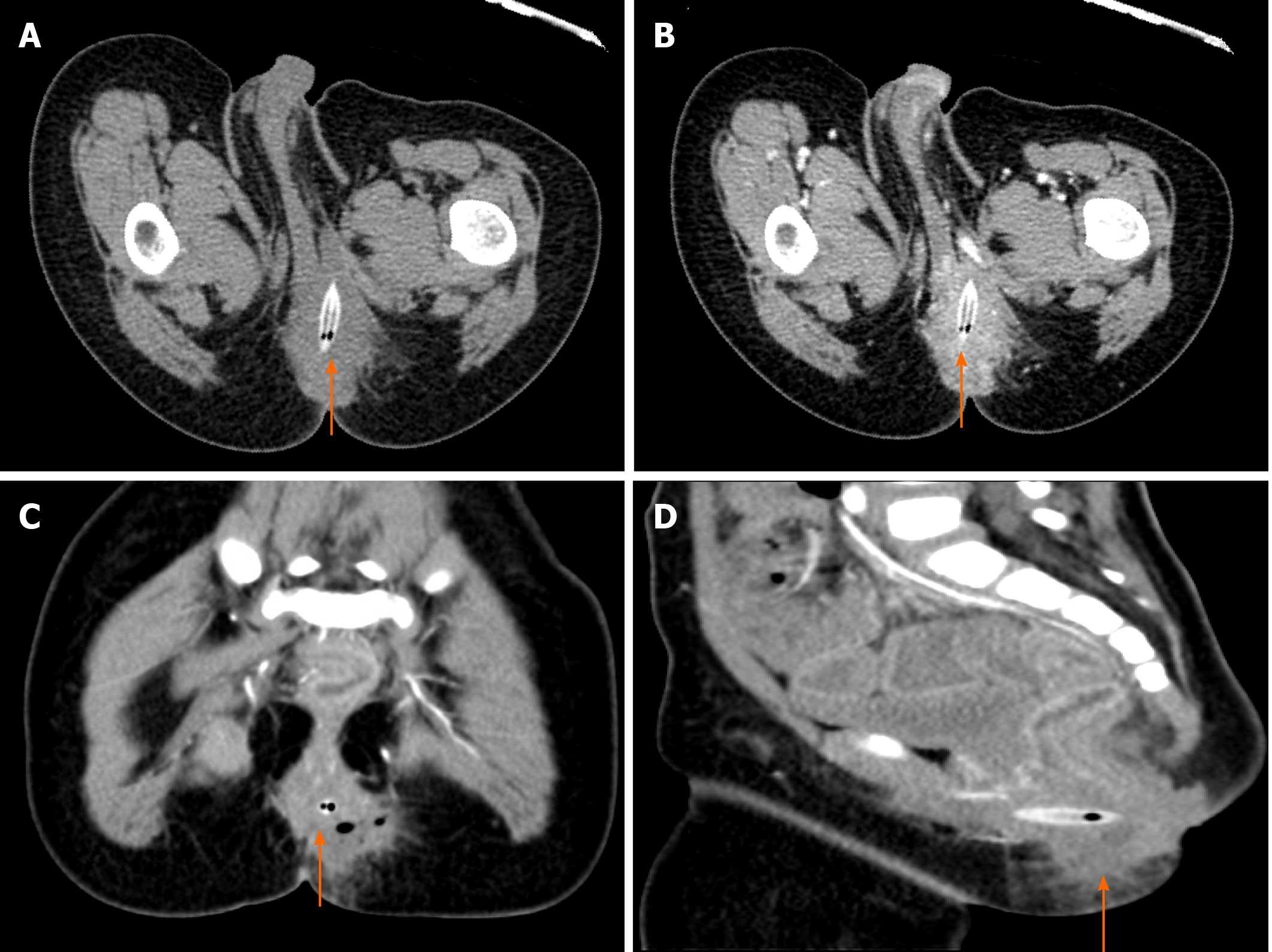
Figure 2 Preoperative abdominal computed tomography scan with intravenous contrast media administration of patient 2.
Axial view (A) and intravenous contrast-enhanced axial (B), coronal (C) and sagittal (D) views showing a foreign body (arrowheads) with mixed density and brim enhancement.
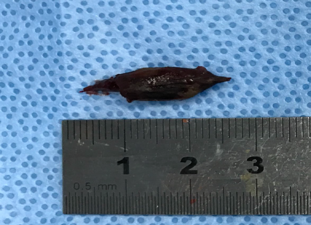
Figure 3 The jujube pit embedded within the fistula in patient 1.
Figure 4 Clinical timeline of patient 1.
FB: Foreign body.
Figure 5 Clinical timeline of patient 2.
FB: Foreign body.
- Citation: Liu YH, Lv ZB, Liu JB, Sheng QF. Perianorectal abscesses and fistula due to ingested jujube pit in infant: Two case reports. World J Clin Cases 2020; 8(20): 4930-4937
- URL: https://www.wjgnet.com/2307-8960/full/v8/i20/4930.htm
- DOI: https://dx.doi.org/10.12998/wjcc.v8.i20.4930


