Copyright
©The Author(s) 2020.
World J Clin Cases. Oct 26, 2020; 8(20): 4917-4921
Published online Oct 26, 2020. doi: 10.12998/wjcc.v8.i20.4917
Published online Oct 26, 2020. doi: 10.12998/wjcc.v8.i20.4917
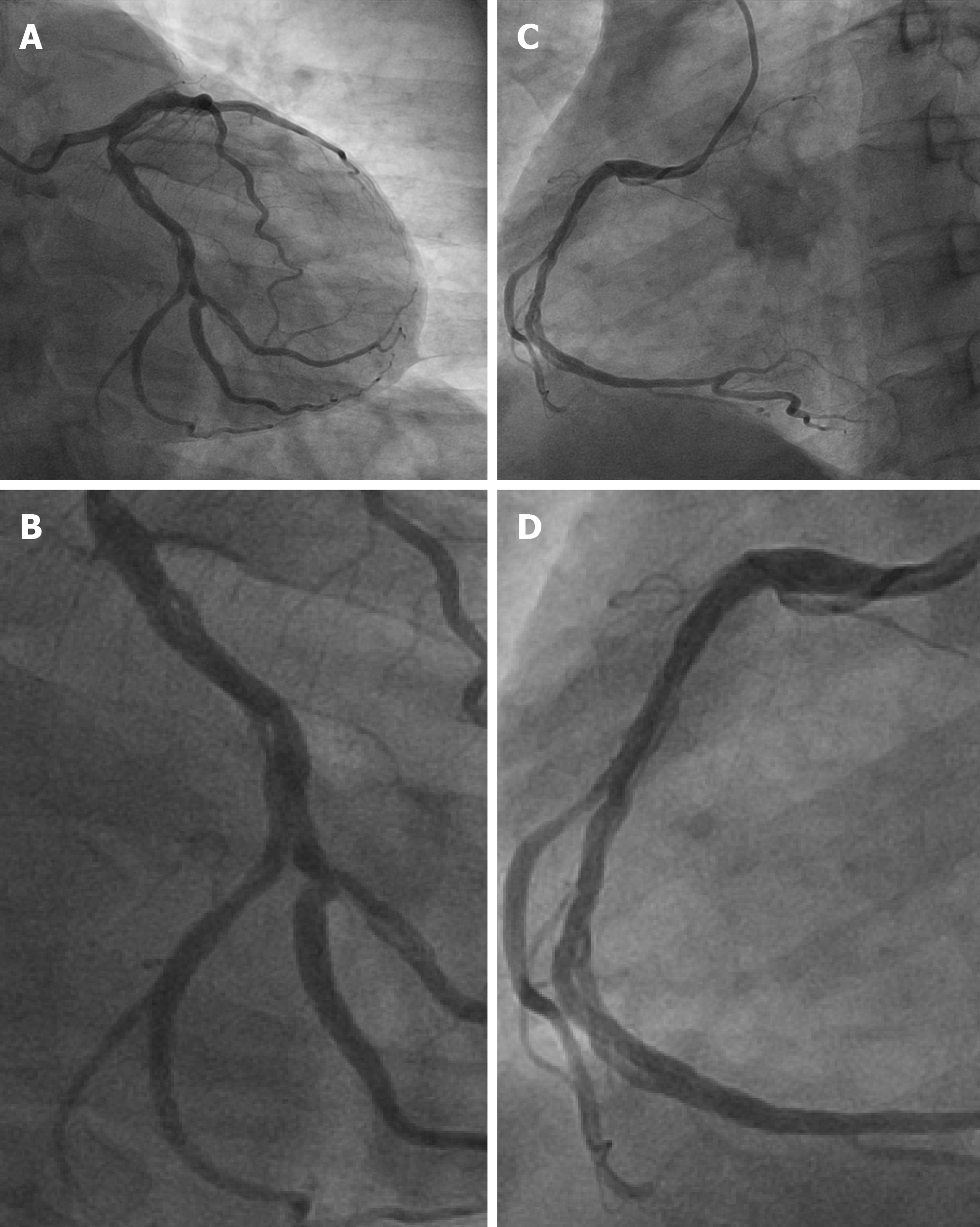
Figure 1 Image of the coronary angiography.
A and B: The “woven” change in left circumflex; C and D: The “woven” change in right coronary artery.
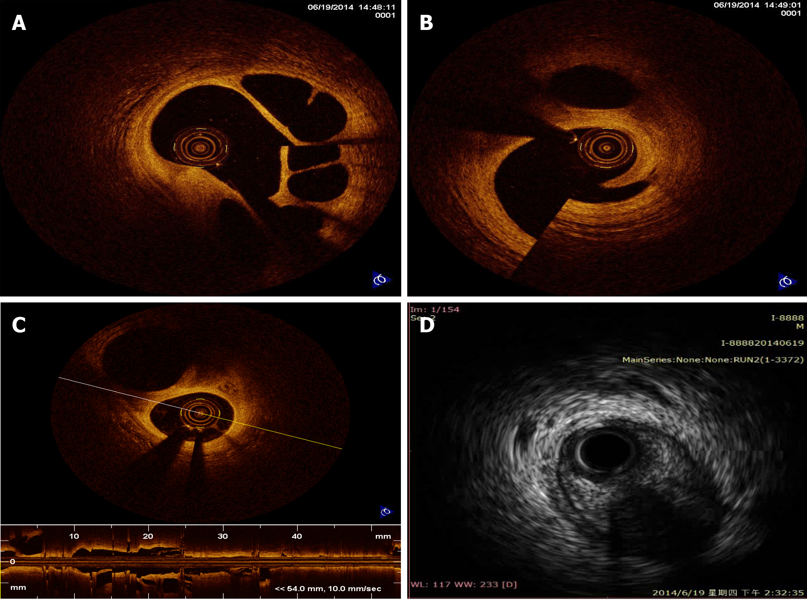
Figure 2 Optical coherence tomography and intravascular ultrasound images in right coronary artery.
A-C: Multiple twisted channels without traces of thrombosis or dissection flaps are shown by optical coherence tomography in right coronary artery; D: Intravascular ultrasound shows multiple cavities filled with blood speckling in right coronary artery.
- Citation: Wei W, Zhang Q, Gao LM. Woven coronary artery: A case report. World J Clin Cases 2020; 8(20): 4917-4921
- URL: https://www.wjgnet.com/2307-8960/full/v8/i20/4917.htm
- DOI: https://dx.doi.org/10.12998/wjcc.v8.i20.4917


