Copyright
©The Author(s) 2020.
World J Clin Cases. Oct 26, 2020; 8(20): 4866-4875
Published online Oct 26, 2020. doi: 10.12998/wjcc.v8.i20.4866
Published online Oct 26, 2020. doi: 10.12998/wjcc.v8.i20.4866
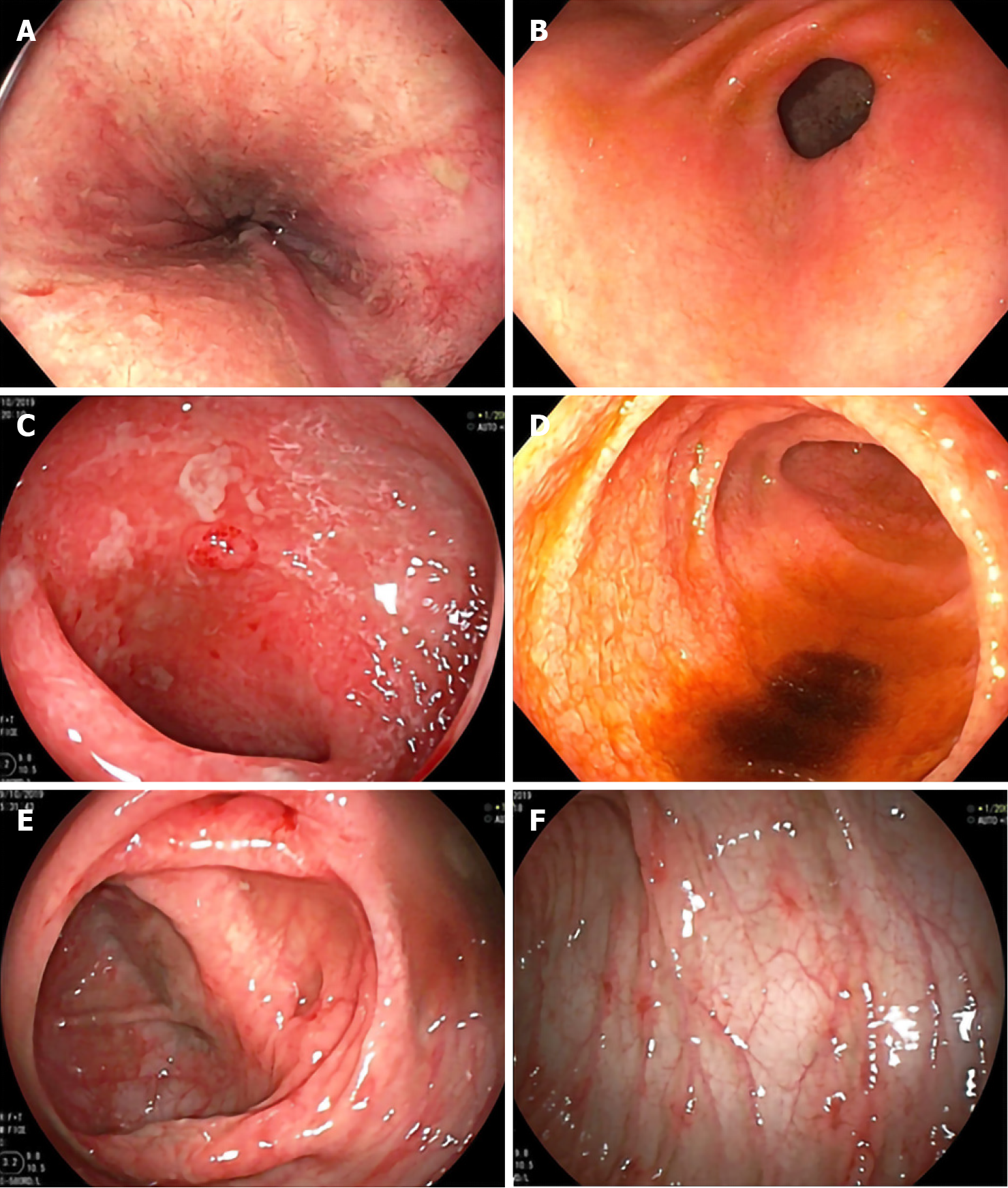
Figure 1 Esophagogastroduodenoscopy and colonoscopy demonstrated erythaematous mucosae with multiple shallow ulcers and yellowish debris scattering along gastrointestinal tract.
A: Lower oesophagus; B: Antrum; C: Duodenum; D: Terminal ileum; E: Transverse colon (hepatic flexure area); F: Descending colon.
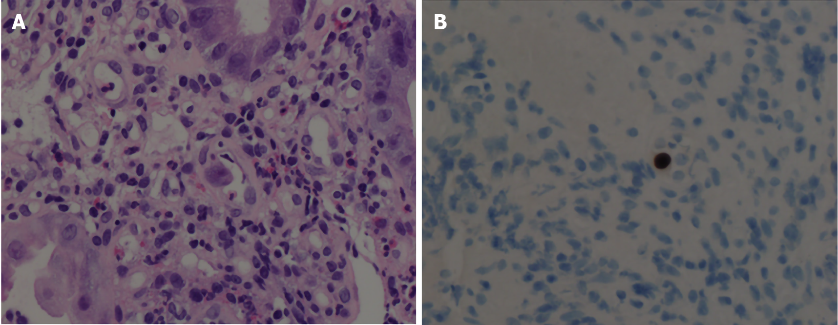
Figure 2 Histopathology demonstrates viral-infected cells.
A: Hematoxylin and eosin stain of colonic tissue showed viral-infected cells (arrow) and mixed inflammatory cell infiltrate in lamina propria; B: Cytomegalovirus immunohistochemistry is positive.
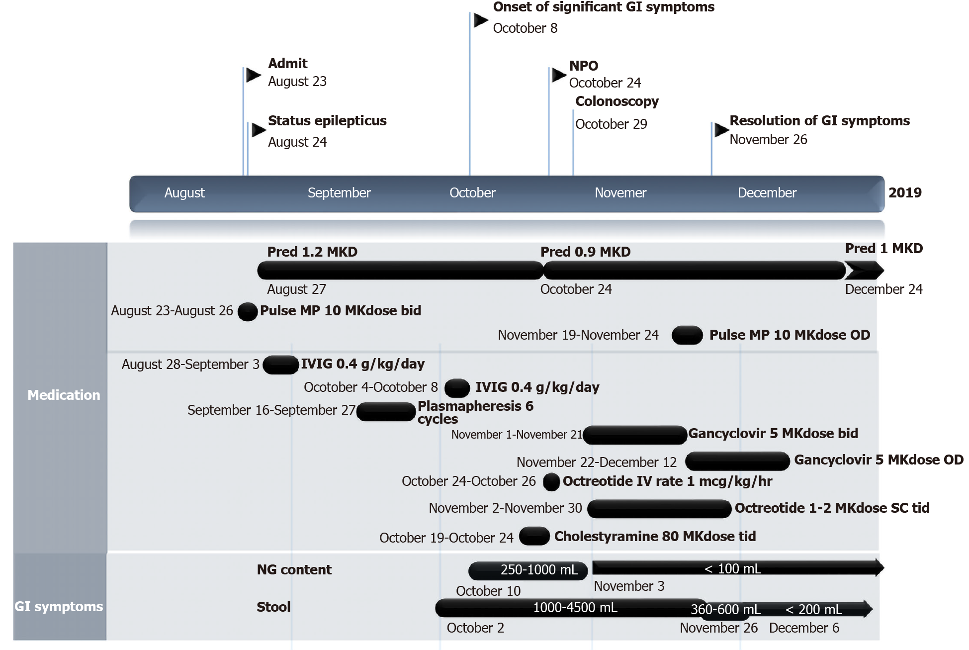
Figure 3 Clinical course of patient since the anti-N-methyl-D-aspartate-receptor encephalitis was diagnosed until severe gastrointestinal symptoms subsided.
NPO: Nil per os; GI: Gastrointestinal; Pred: Prednisolone; MP: Methylprednisolone; IVIG: Intravenous immunoglobulin; MKdose: mg per kg per dose; MKD: mg per kg per day; IV: Intravenous; OD: Once a day; bid: Twice a day; tid: Three times a day; NG: Nasogastric; ALC: Absolute lymphocyte count; CMV: Cytomegalovirus.
- Citation: Onpoaree N, Veeravigrom M, Sanpavat A, Suratannon N, Sintusek P. Unremitting diarrhoea in a girl diagnosed anti-N-methyl-D-aspartate-receptor encephalitis: A case report. World J Clin Cases 2020; 8(20): 4866-4875
- URL: https://www.wjgnet.com/2307-8960/full/v8/i20/4866.htm
- DOI: https://dx.doi.org/10.12998/wjcc.v8.i20.4866


