©The Author(s) 2020.
World J Clin Cases. Oct 6, 2020; 8(19): 4681-4687
Published online Oct 6, 2020. doi: 10.12998/wjcc.v8.i19.4681
Published online Oct 6, 2020. doi: 10.12998/wjcc.v8.i19.4681
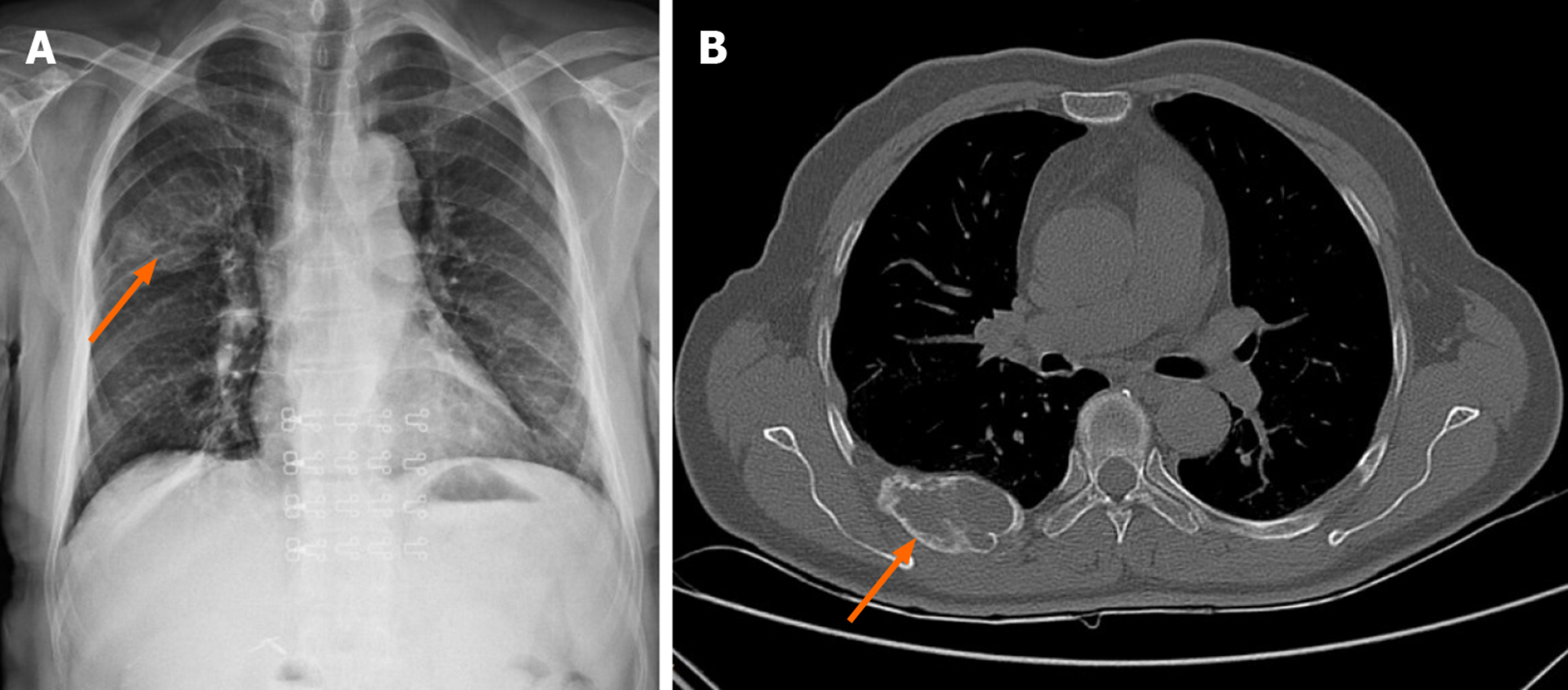
Figure 1 Chest X-ray and computed tomography before surgery.
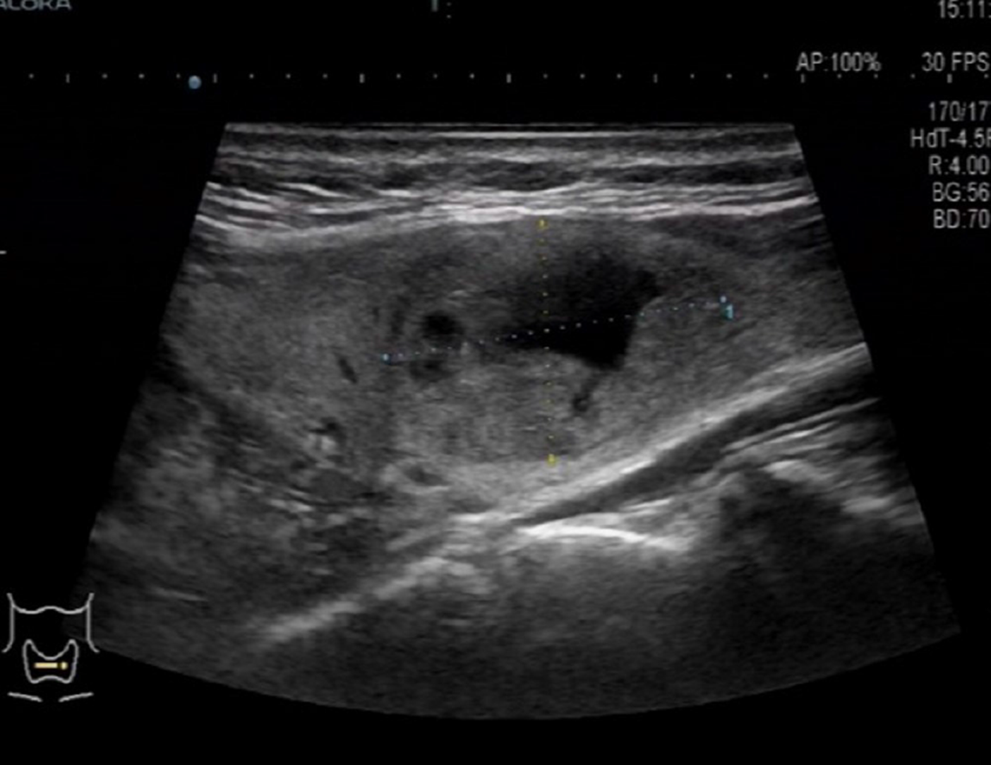
Figure 2 Ultrasound image of the thyroid.
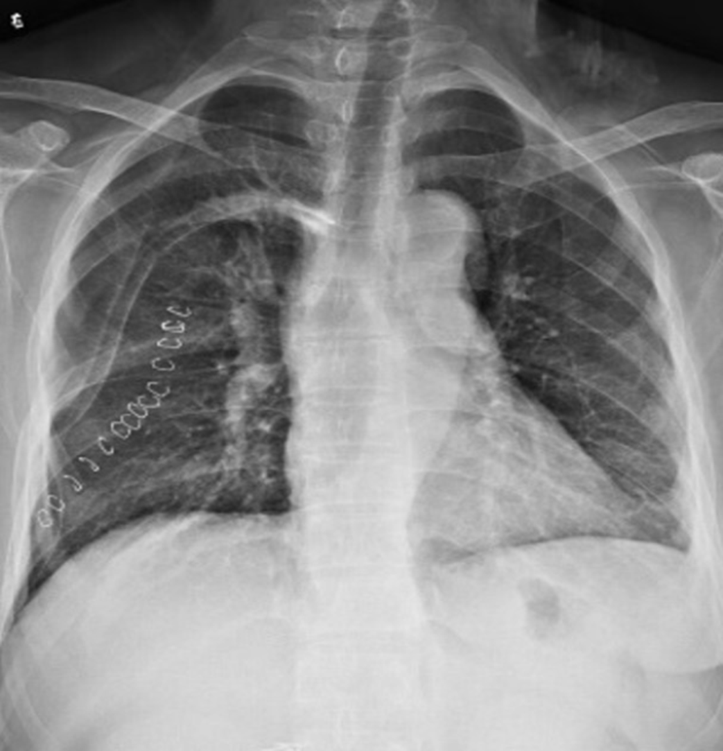
Figure 3 Chest X-ray after surgery.
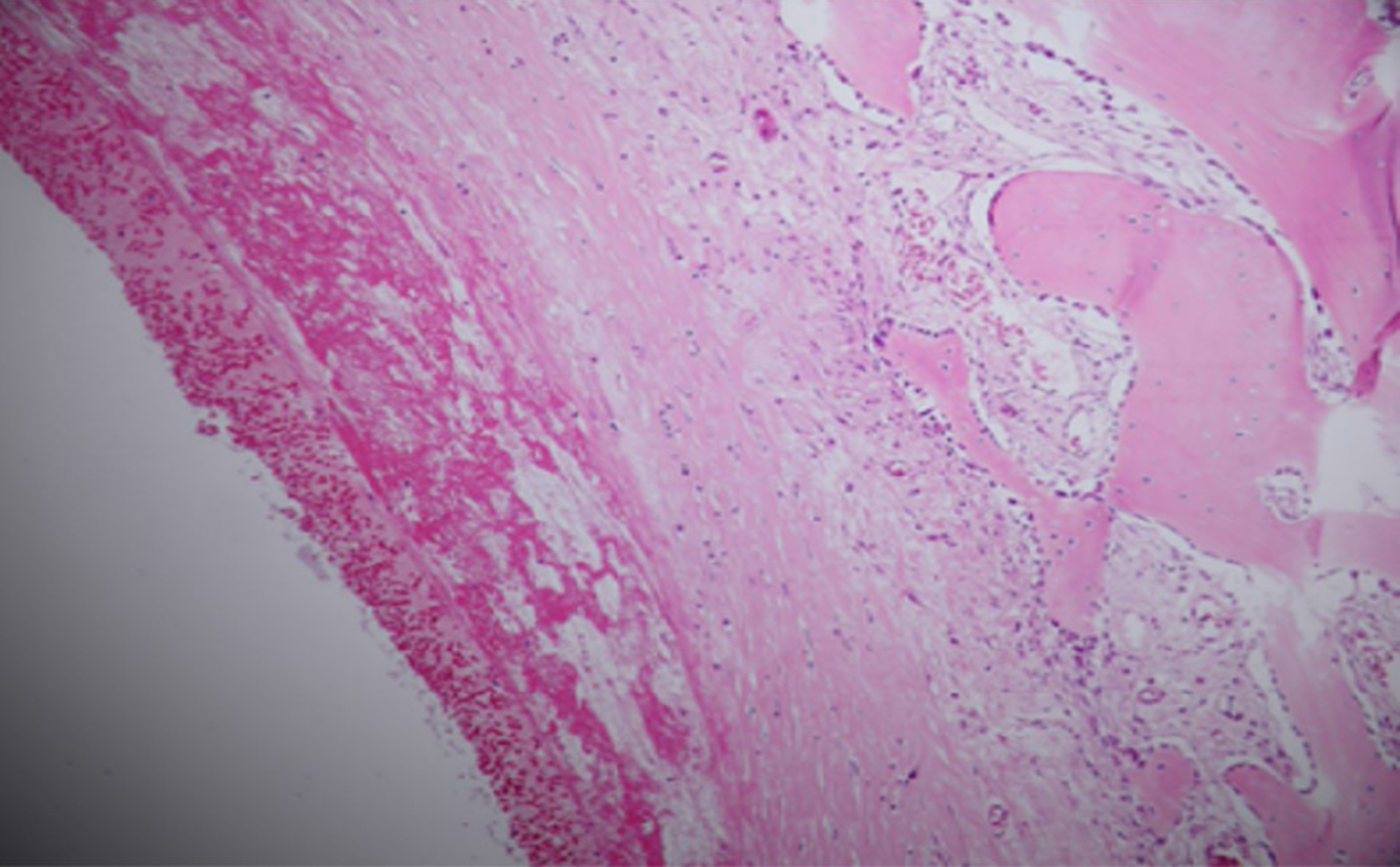
Figure 4 Pathological results of the rib tumor.
A solid section of the cut surface, bleeding and bloody fluid are seen in the cyst cavity, fibrous tissue hyperplasia around the cystic cavity, reactive hyperplasia of bone-like tissue and trabecular bone in the deep cystic cavity, solid part of the area containing hemosiderin deposition, and multinucleated giant cell reaction.
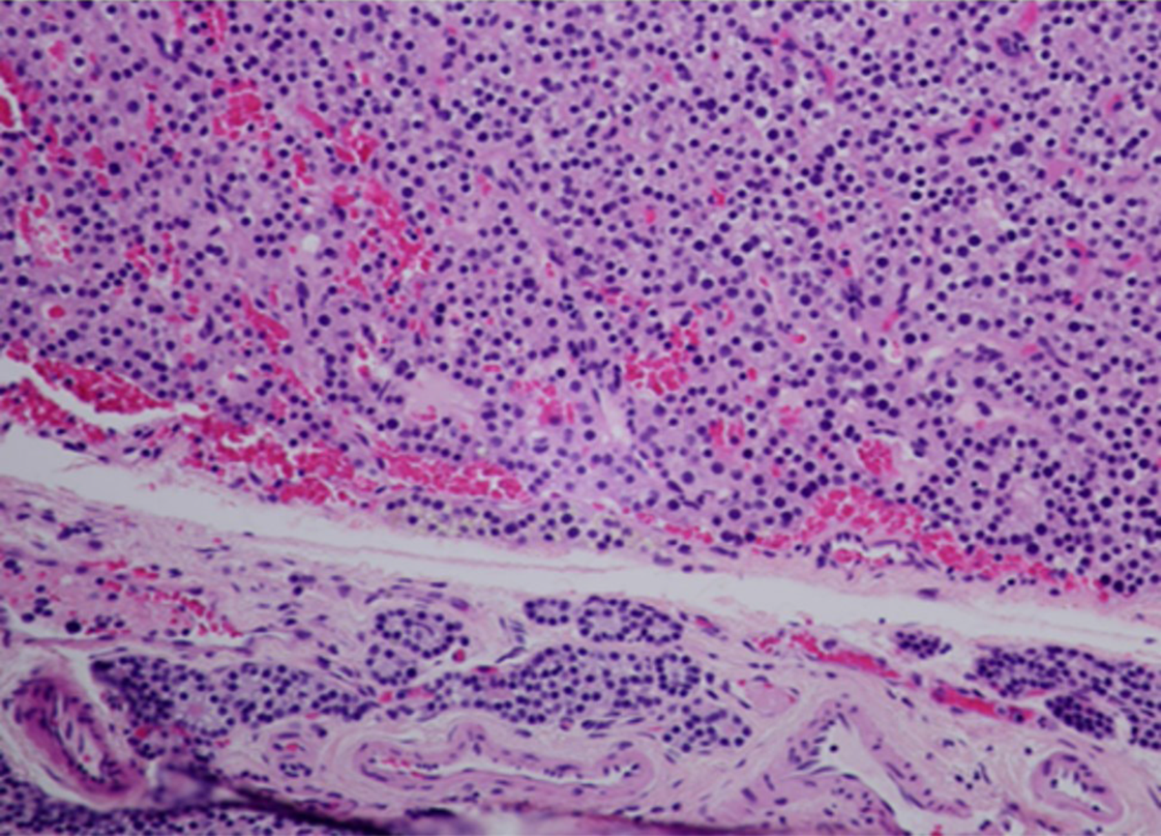
Figure 5 Pathological results of the parathyroid adenoma.
Left lower parathyroid gland combined with HE morphology and immunohistochemistry results diagnosis: parathyroid adenoma. Immunohistochemical results: CgA part (+), PTH (+), Ki67 (marker of proliferation Ki-67) approximately 1% (+), TG (-), and TTF-1 (thyroid transcription factor 1) (-).
- Citation: Han L, Zhu XF. Parathyroid adenoma combined with a rib tumor as the primary disease: A case report. World J Clin Cases 2020; 8(19): 4681-4687
- URL: https://www.wjgnet.com/2307-8960/full/v8/i19/4681.htm
- DOI: https://dx.doi.org/10.12998/wjcc.v8.i19.4681


