©The Author(s) 2020.
World J Clin Cases. Oct 6, 2020; 8(19): 4488-4493
Published online Oct 6, 2020. doi: 10.12998/wjcc.v8.i19.4488
Published online Oct 6, 2020. doi: 10.12998/wjcc.v8.i19.4488
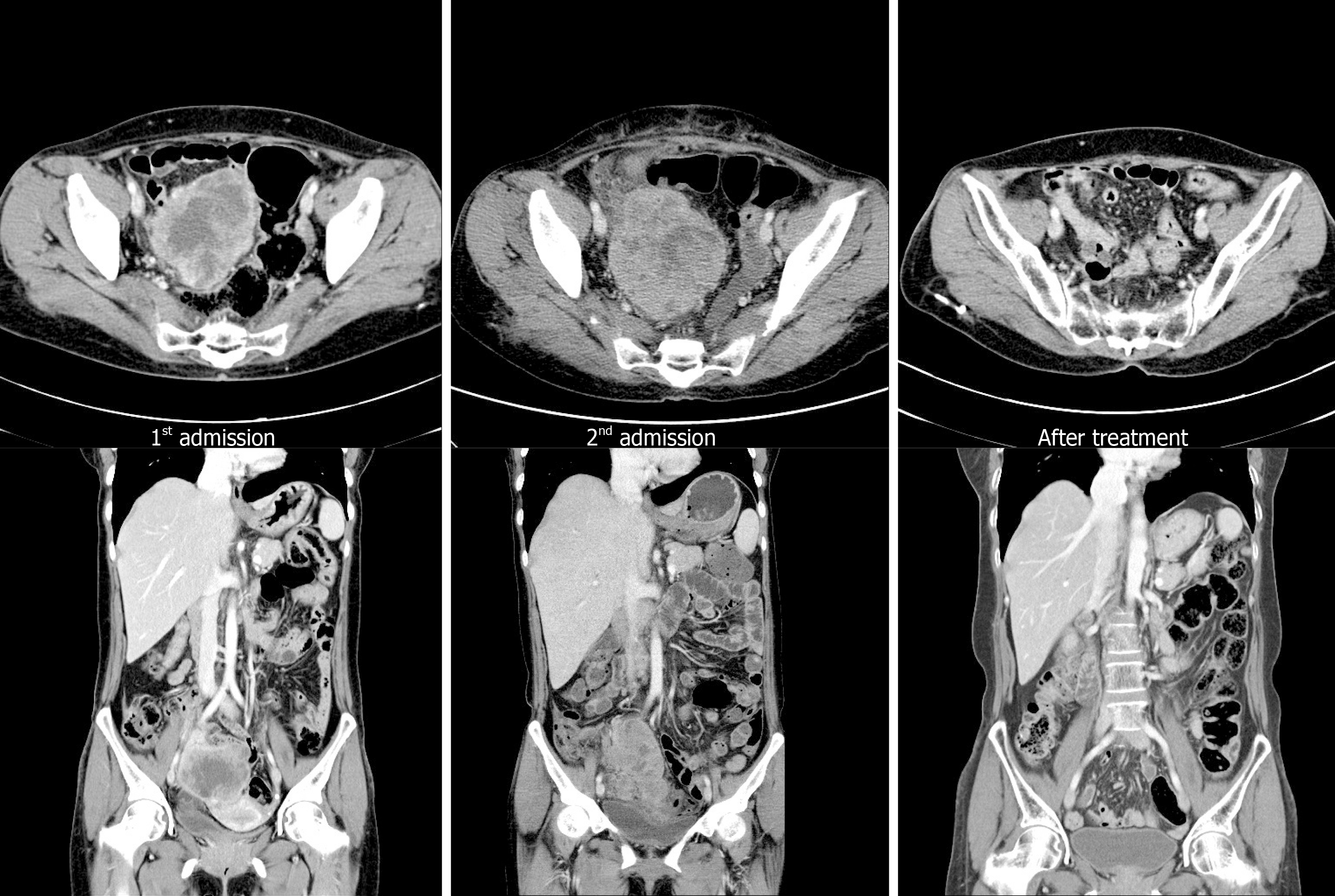
Figure 1 Computed tomography at admission and follow-up.
Computed tomography (CT) scan performed at first admission showing an 8 cm × 7 cm size mass only in the pelvic cavity. However, 6 wk after surgery (second admission), CT scan shows a mass that was larger than the initial mass in the pelvic cavity with peritoneal seeding and para-aortic lymphadenopathy (arrow). These lesions are almost no longer observed after chemotherapy.
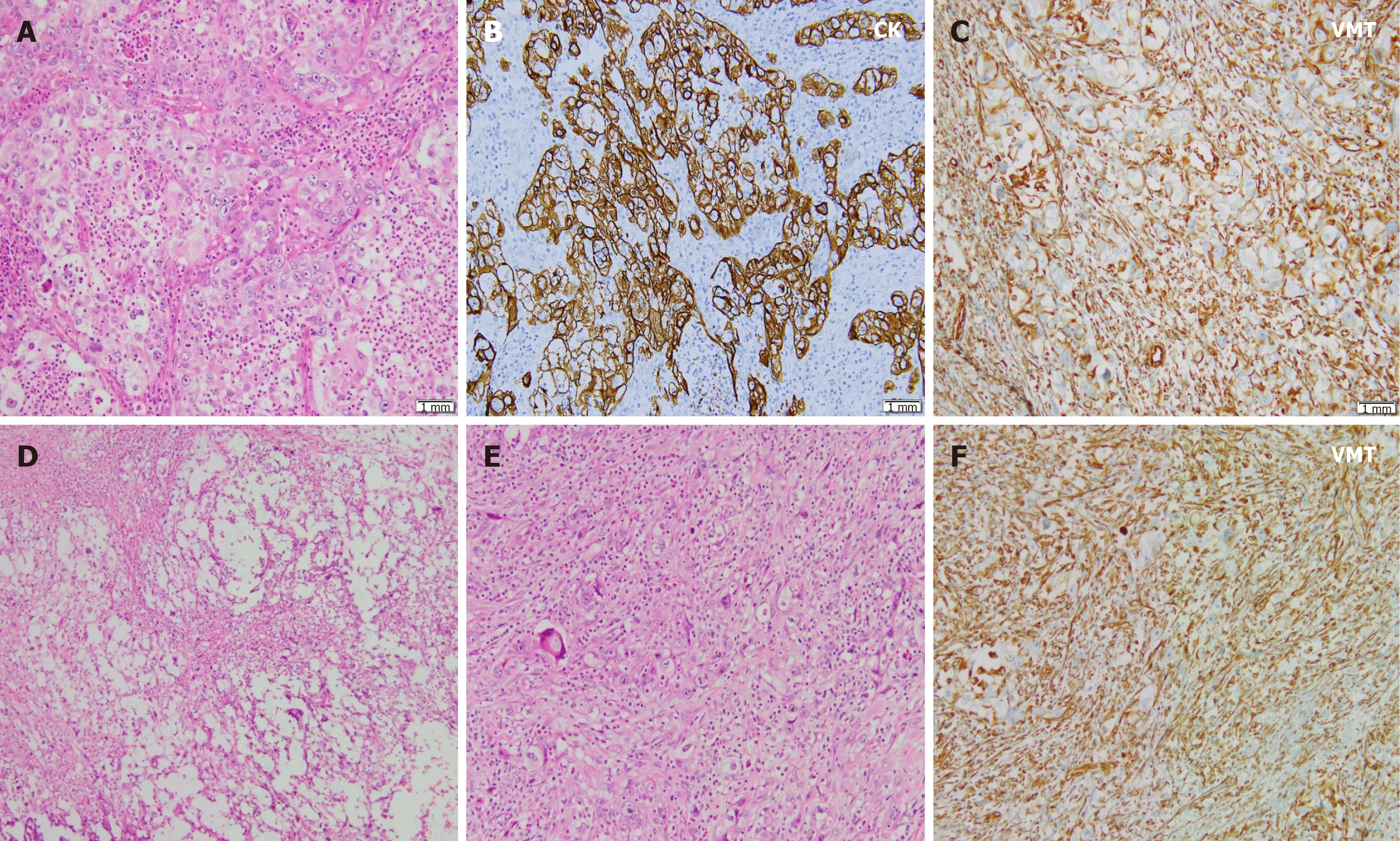
Figure 2 Pathologic findings.
A: Tumor cell nests such as round to oval nuclei, prominent nucleoli, and many atypical mitotic figures are observed in the neutrophilic background; B and C: These cells are immunoreactive to pan-cytokeratin, but negative for vimentin (VMT) (B) and focally to VMT (C); D: Geographic necrosis is common with myxoid matrix; E: In some areas, sarcomatoid features are frequently observed; F: Tumor cells showing no organoid feature directly admixed with stromal components and were immunoreactive to VMT. CK: Cytokeratin; VMT: Vimentin.
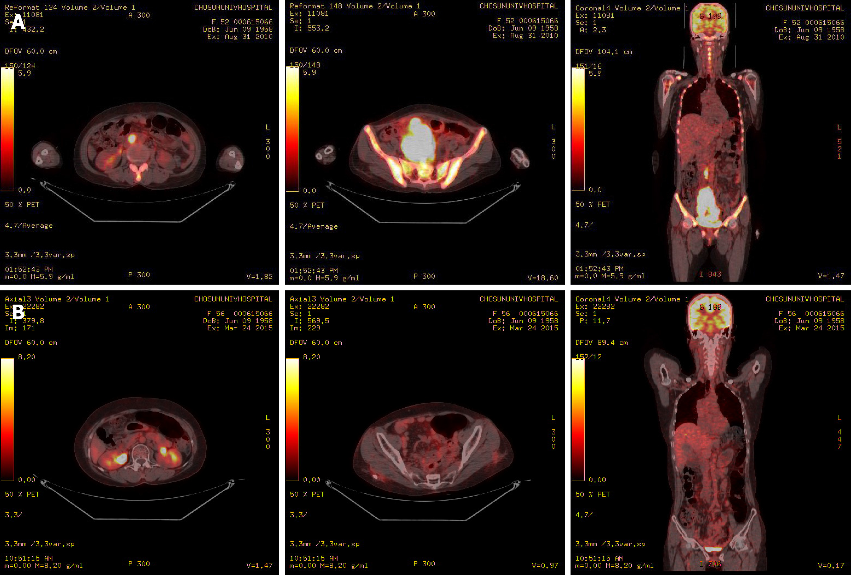
Figure 3 Positron emission tomography–computed tomography.
A: Positron emission tomography–computed tomography revealing hypermetabolism of the huge mass in the pelvic cavity, multiple peritoneal nodules, and para-aortic lymph nodes; B: Hypermetabolism of the huge mass in the pelvic cavity, multiple peritoneal nodules, and para-aortic lymph nodes showing improvement after chemotherapy.
- Citation: Kim HB, Lee HJ, Hong R, Park SG. Extremely rare case of successful treatment of metastatic ovarian undifferentiated carcinoma with high-dose combination cytotoxic chemotherapy: A case report. World J Clin Cases 2020; 8(19): 4488-4493
- URL: https://www.wjgnet.com/2307-8960/full/v8/i19/4488.htm
- DOI: https://dx.doi.org/10.12998/wjcc.v8.i19.4488


