©The Author(s) 2020.
World J Clin Cases. Sep 26, 2020; 8(18): 4215-4222
Published online Sep 26, 2020. doi: 10.12998/wjcc.v8.i18.4215
Published online Sep 26, 2020. doi: 10.12998/wjcc.v8.i18.4215
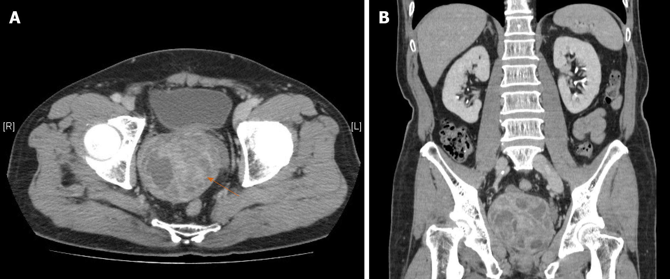
Figure 1 Enhanced computed tomography of the abdomen and pelvis, (A) axial and (B) coronal views, showing a large multiloculated mass with heterogeneous density (arrow) arising from the prostate gland and measuring about 8.
2 cm × 8.1 cm × 7.8 cm in size. Although the tumor occupied almost all the pelvic space, it adhered to the surrounding structures without invasion of the bladder or rectum.
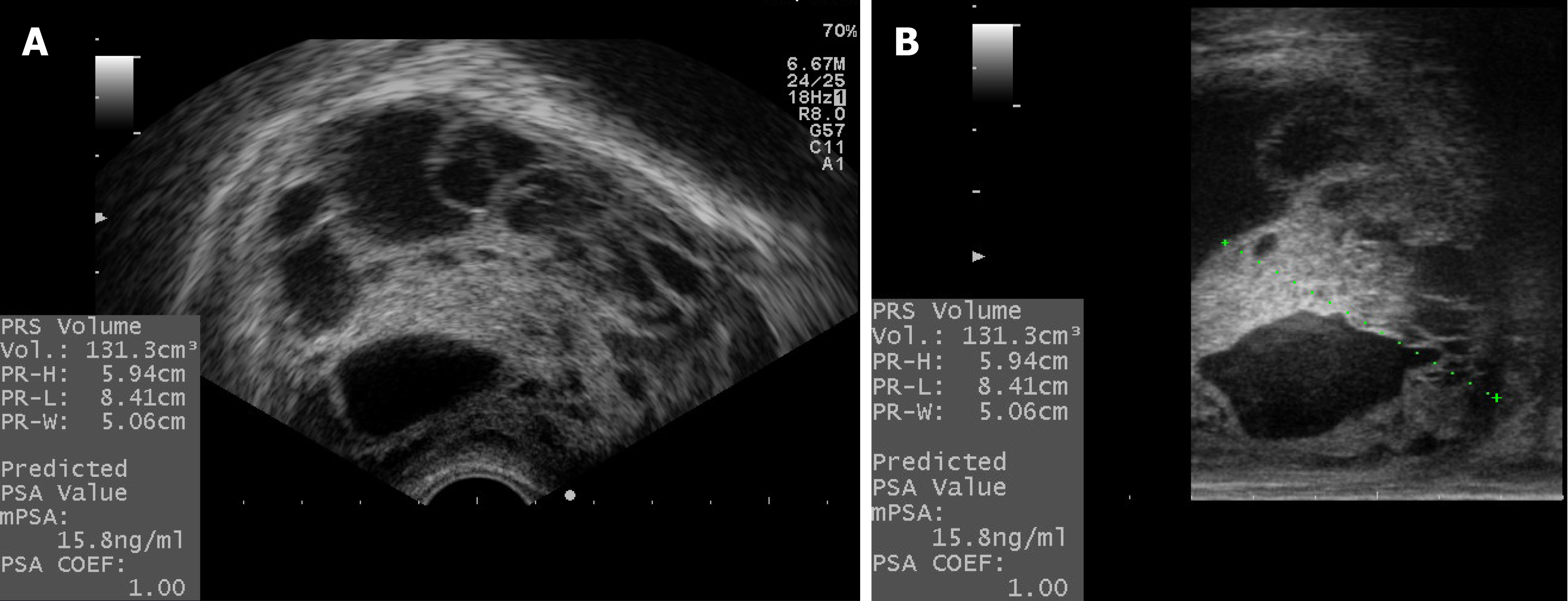
Figure 2 Transrectal ultrasound revealed a huge prostate gland measuring 8.
41 cm × 5.94 cm × 5.06 cm, with a corresponding volume of 131.3 mL, with heterogeneous echoic density and multiple loculated irregular cysts embedded in the prostate gland, which could not be distinguished from the junction of the transitional and peripheral zones regardless of the view, (A) transverse or (B) sagittal.
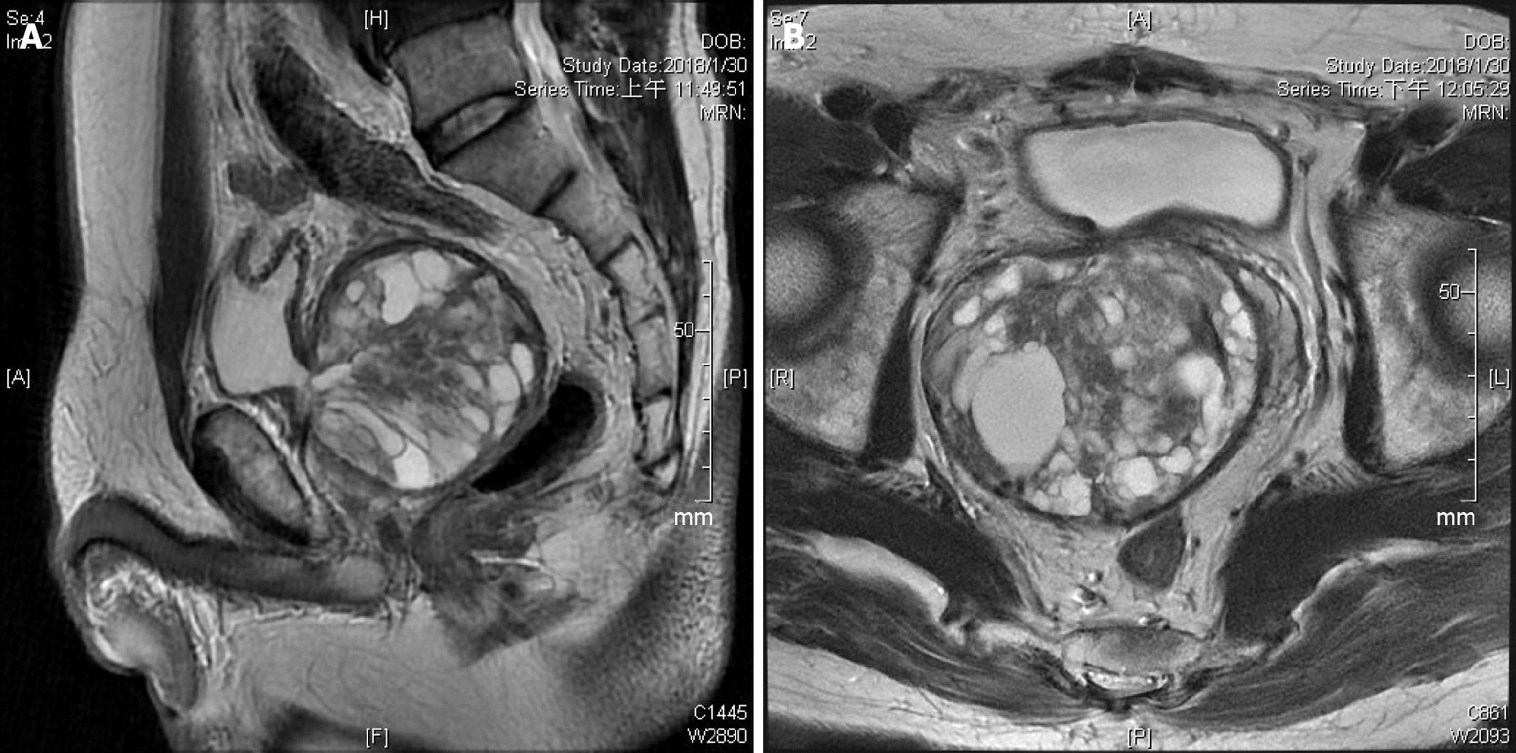
Figure 3 T2-weighted magnetic resonance imaging showing a recurrent cystic multiseptate mass displacing the surrounding structures, (A) sagittal and (B) axial views.
Heterogeneous signal intensity suggests simple fluid, fat, or blood in the cysts. Because of some solid components with low signal intensity in the central part of the tumor, the possibility of a co-existing malignancy could not be eliminated.
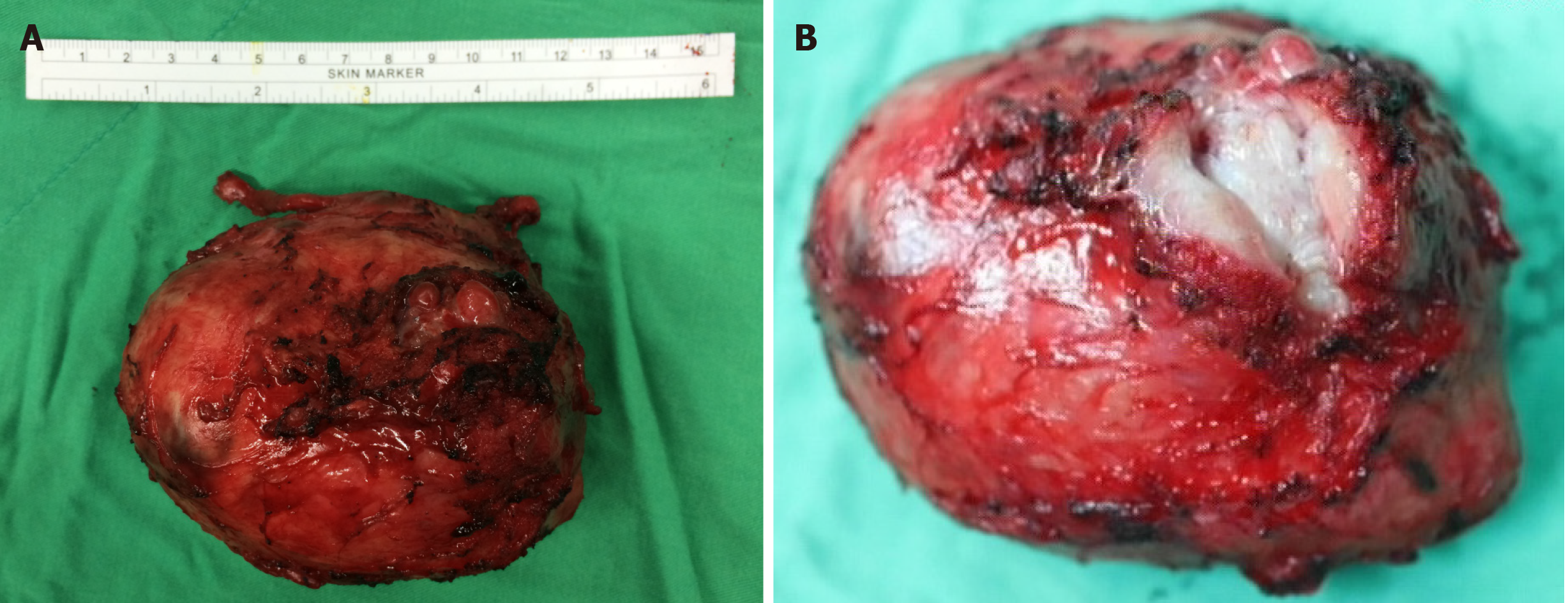
Figure 4 Gross photographs.
A: Prostate gland with bilateral seminal vesicles resected en bloc, measuring 11.1 cm × 8.2 cm × 7.1 cm in size, The asterisk marks the bladder neck, which shows regrowth of the adenoma from the transition zone and which had undergone TURP previously; B: Anterior view of prostate incised to show narrowing of the prostate urethra, even after 3 transurethral prostatectomies. The whole tumor was removed intact, without rupture of the cystic components. The tumor protrudes from the prostate gland posteriorly and compresses the bilateral vas deferens, resulting in dilated seminal vesicles.
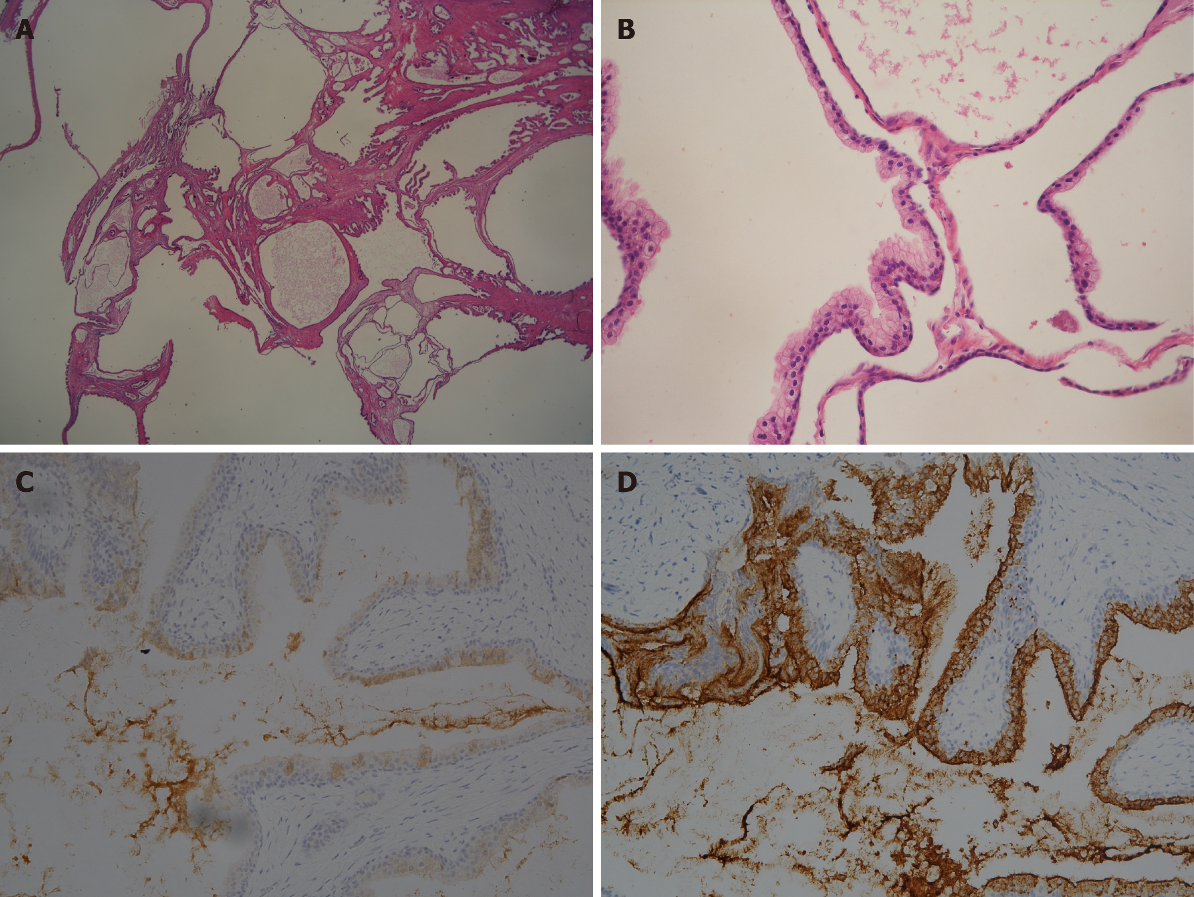
Figure 5 Microscopic findings of the resected tissue.
A: Variously sized dilated glandular and cystic structures lined by blended prostatic type epithelia on low-power magnification (hematoxylin and eosin, original magnification 10 ×); B: Glands and cysts are lined by cuboidal to columnar epithelial cells with a distinct, intact basal cell layer (40 ×); C and D: The epithelium of the cysts stained positively for prostate-specific antigen and weakly expressed prostate-specific membrane antigen (20 ×).
Figure 6 Timeline of the treatment.
TURP: Transurethral resections of the prostate; RARP: Robot-assisted radical prostatectomy.
- Citation: Fan LW, Chang YH, Shao IH, Wu KF, Pang ST. Robotic surgery in giant multilocular cystadenoma of the prostate: A rare case report. World J Clin Cases 2020; 8(18): 4215-4222
- URL: https://www.wjgnet.com/2307-8960/full/v8/i18/4215.htm
- DOI: https://dx.doi.org/10.12998/wjcc.v8.i18.4215


