©The Author(s) 2020.
World J Clin Cases. Sep 26, 2020; 8(18): 4207-4214
Published online Sep 26, 2020. doi: 10.12998/wjcc.v8.i18.4207
Published online Sep 26, 2020. doi: 10.12998/wjcc.v8.i18.4207
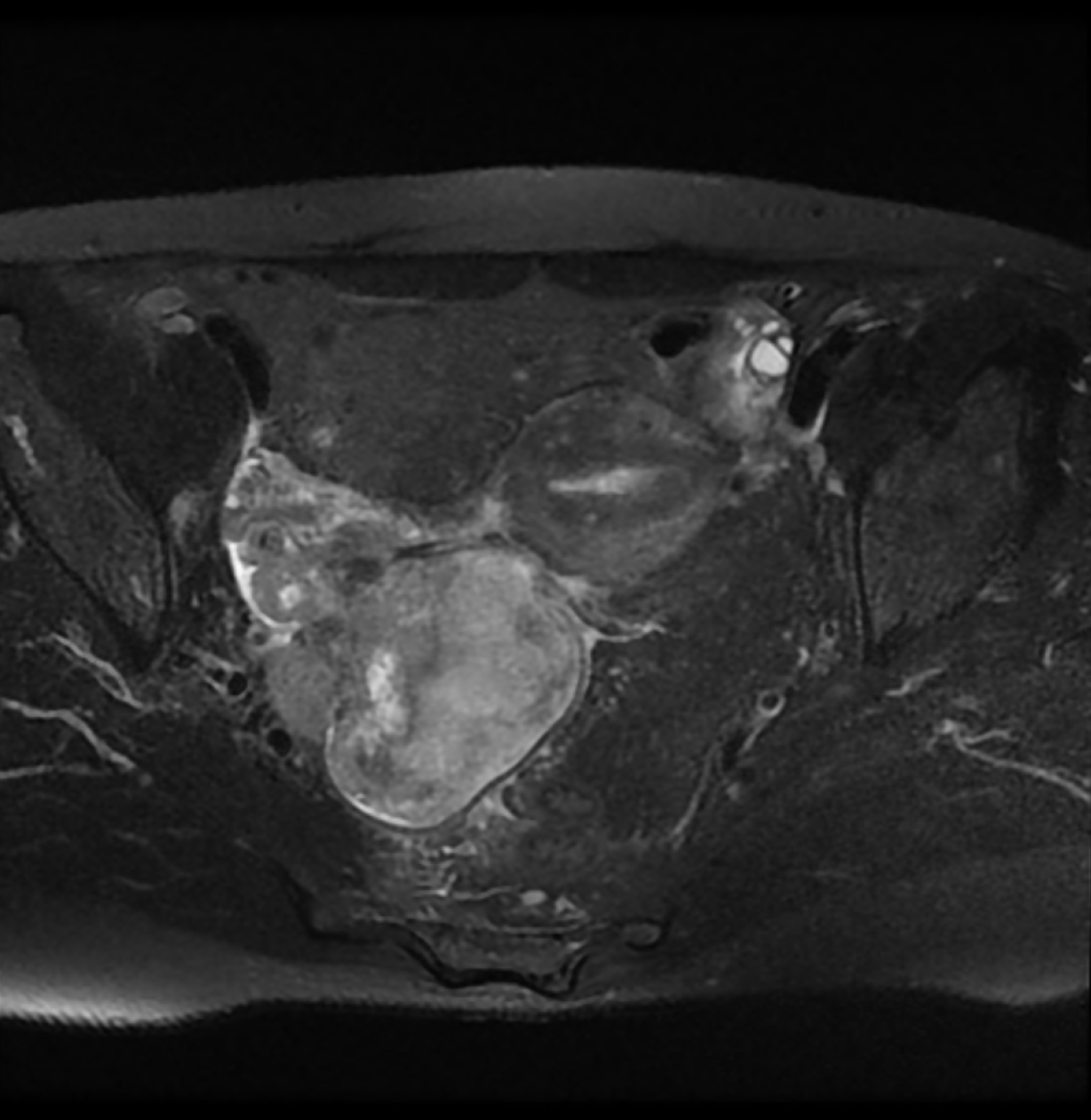
Figure 1 Pelvis magnetic resonance imaging showing heterogeneously enhancing mass (8 cm × 6 cm × 6 cm) occupying right pelvic cavity.
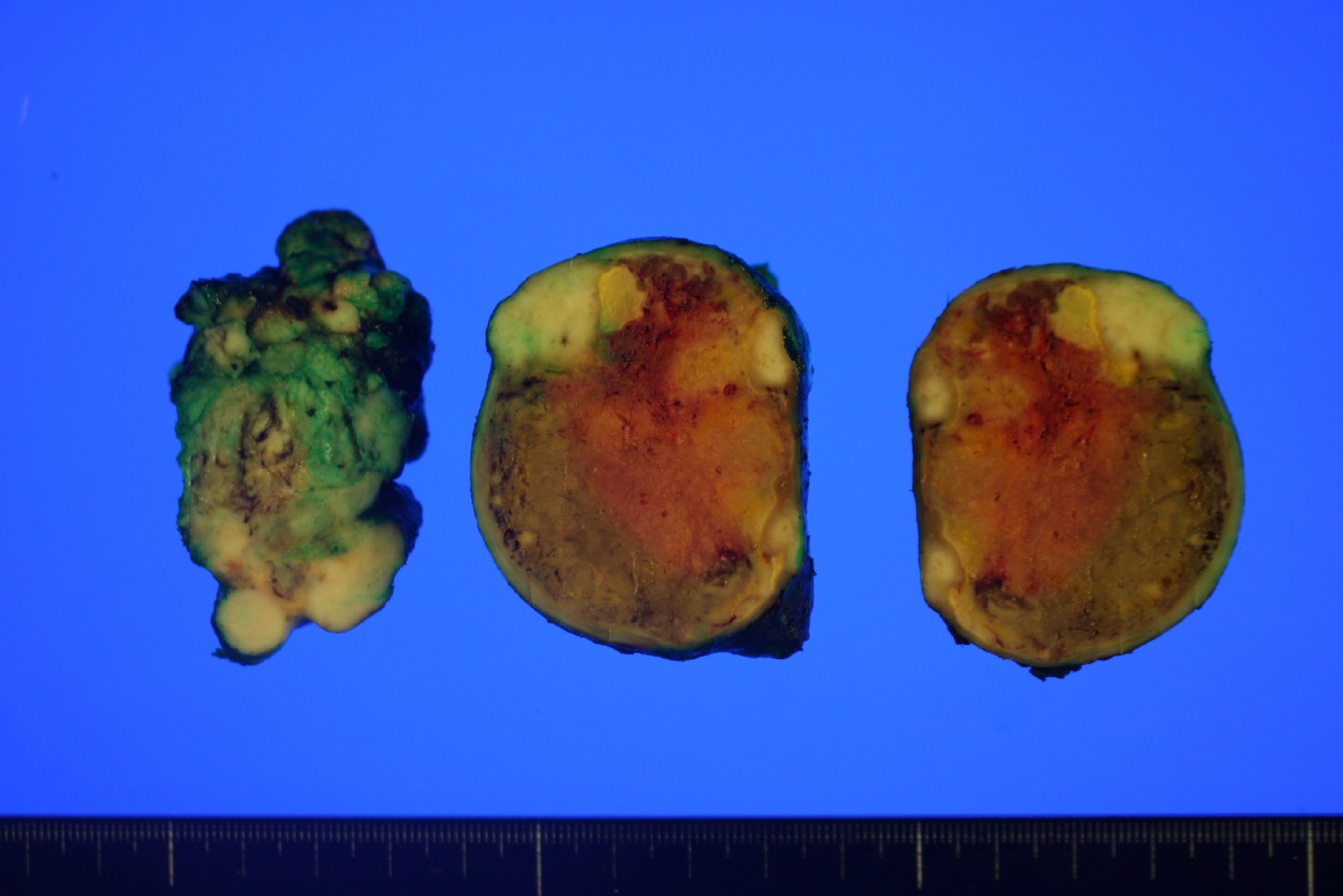
Figure 2 Gross findings exhibiting a relatively well-circumscribed white to tan solid mass with focal necrosis.
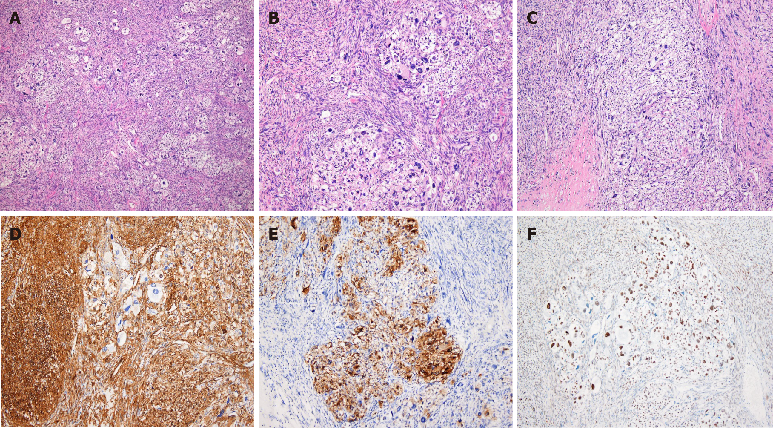
Figure 3 Histopathologic and immunohistochemical findings.
A: The tumor was composed of sheets of epithelioid cells and bundles of spindle cells, hematoxylin and eosin (H&E), × 40; B and C: Mixed epithelioid and spindle cells exhibiting striking pleomorphism and necrosis (H&E, × 100); D: Spindle cells were positive for smooth muscle actin (× 100); E: Epithelioid cells were positive for HMB-45 (× 100); F: Epithelioid cells showed nuclear positivity of TFE3 (× 100).
- Citation: Kim NI, Lee JS, Choi YD, Ju UC, Nam JH. TFE3-expressing malignant perivascular epithelioid cell tumor of the mesentery: A case report and review of literature. World J Clin Cases 2020; 8(18): 4207-4214
- URL: https://www.wjgnet.com/2307-8960/full/v8/i18/4207.htm
- DOI: https://dx.doi.org/10.12998/wjcc.v8.i18.4207


