©The Author(s) 2020.
World J Clin Cases. Sep 26, 2020; 8(18): 4200-4206
Published online Sep 26, 2020. doi: 10.12998/wjcc.v8.i18.4200
Published online Sep 26, 2020. doi: 10.12998/wjcc.v8.i18.4200
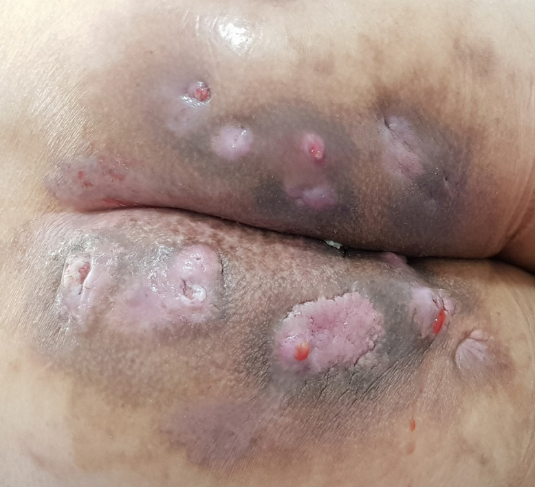
Figure 1 Clinical appearance of buttocks.
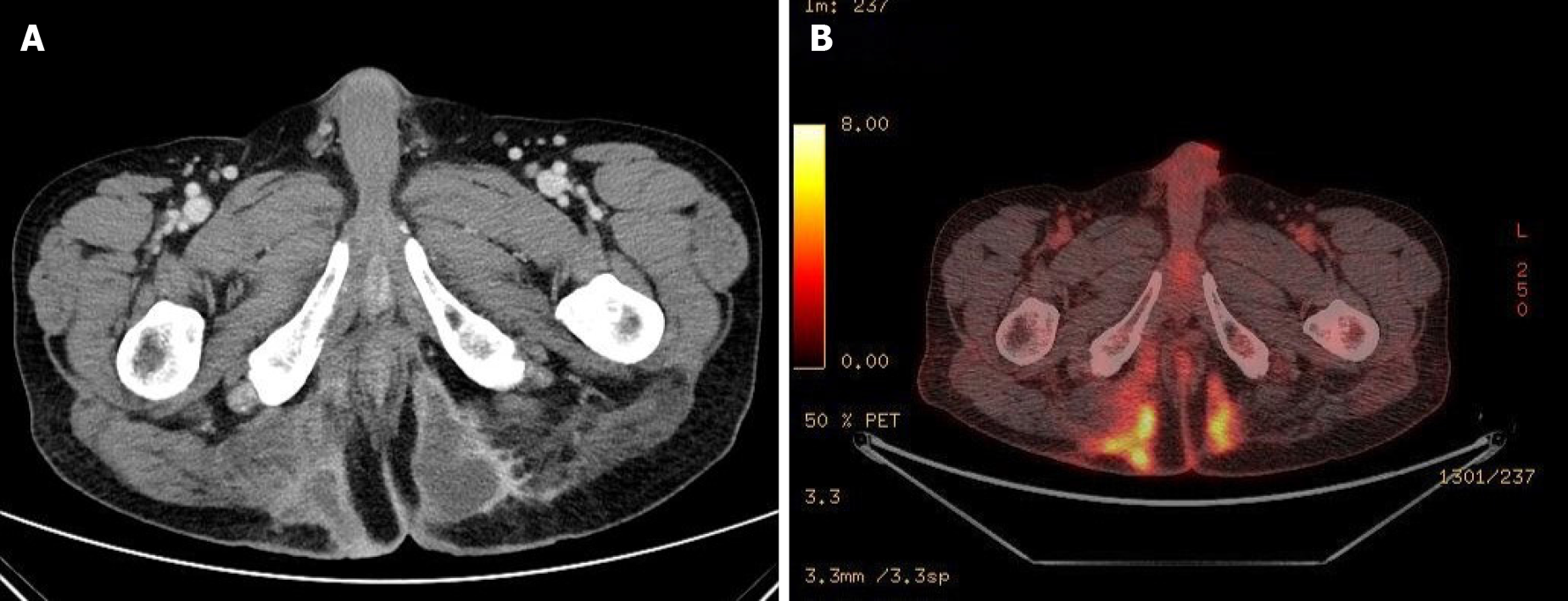
Figure 2 Radiologic findings.
A: Computed tomography findings before the first operation show a huge abscess cavity in bilateral buttocks; B: Positron emission tomography findings show 18F-fluorodeoxyglucose uptake along the abscess cavity.
Figure 3 Pathologic results of the buttock mass.
A: There are variable-sized proliferated atypical glands and mucin pools (H&E, × 4); B: The atypical glands are positive for cytokeratin 20; C: The atypical glands are positive for CDX2.
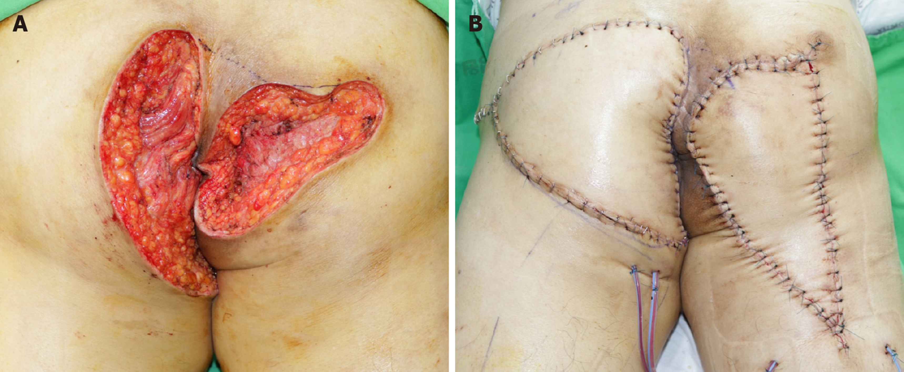
Figure 4 Photographs after the second operation.
A: Wide excision; B: V-Y flap operation.
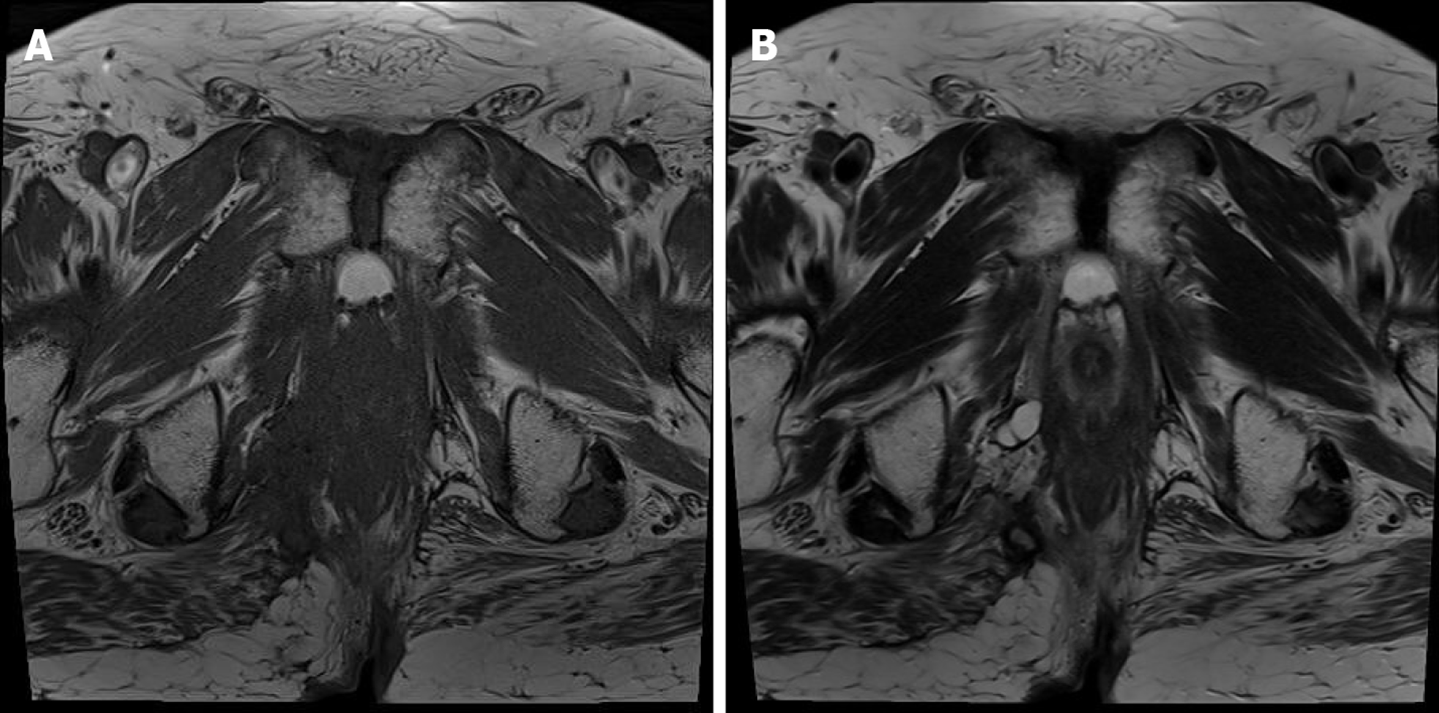
Figure 5 Follow-up magnetic resonance imaging after 24 mo.
There is no recurrence. A: T1 weighted image; B: T2 weighted image.
- Citation: Kim SJ, Kim TG, Gu MJ, Kim S. Mucinous adenocarcinoma of the buttock associated with hidradenitis: A case report. World J Clin Cases 2020; 8(18): 4200-4206
- URL: https://www.wjgnet.com/2307-8960/full/v8/i18/4200.htm
- DOI: https://dx.doi.org/10.12998/wjcc.v8.i18.4200


