Copyright
©The Author(s) 2020.
World J Clin Cases. Sep 26, 2020; 8(18): 4169-4176
Published online Sep 26, 2020. doi: 10.12998/wjcc.v8.i18.4169
Published online Sep 26, 2020. doi: 10.12998/wjcc.v8.i18.4169
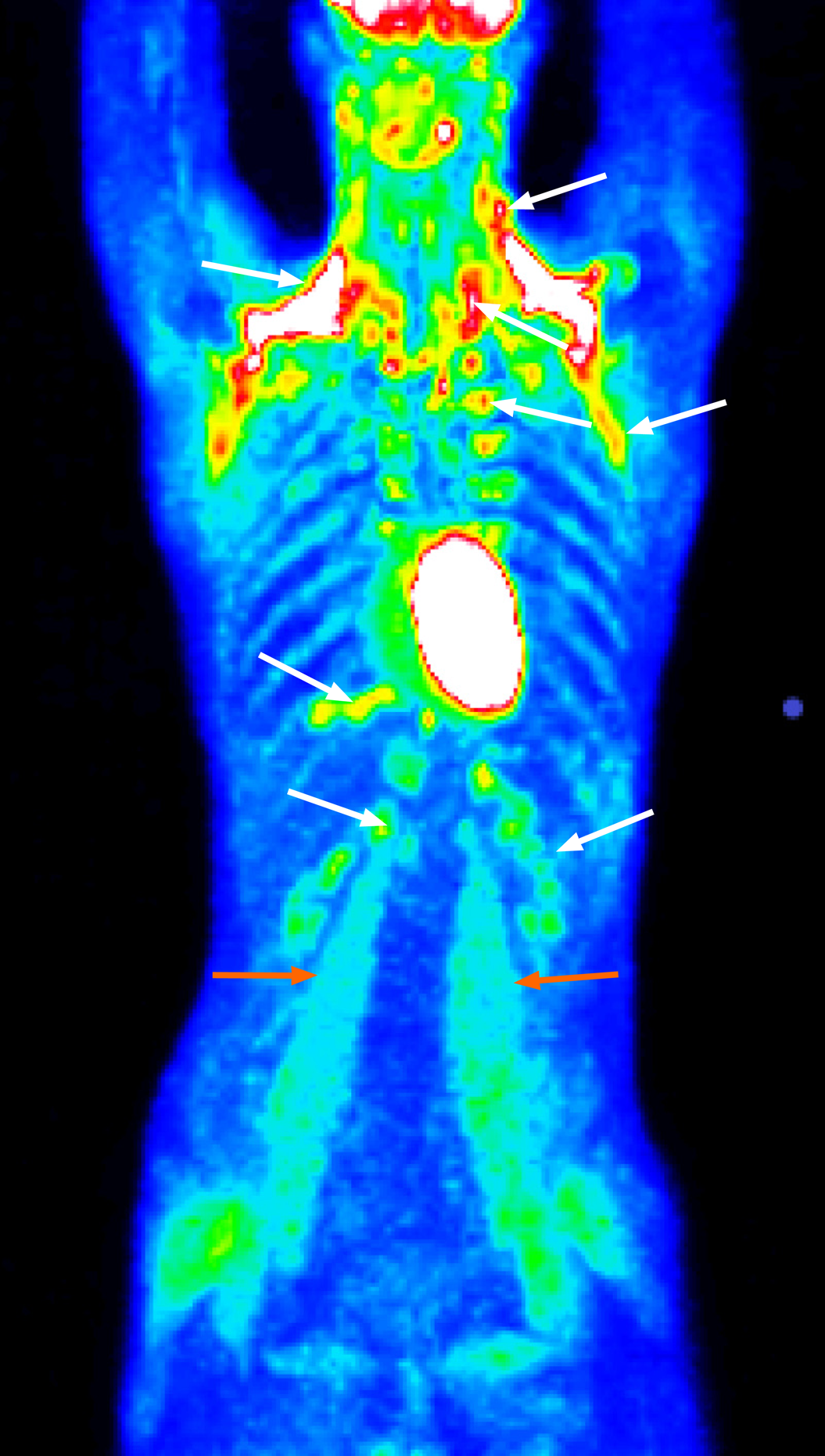
Figure 1 Positron emission tomography with 2-deoxy-2-[fluorine-18]fluoro-D-glucose integrated with computed tomography showing enhanced metabolic activity of brown adipose tissue in the neck, paravertebral, supra- and infraclavicular, axillar, mediastinal, jugular, diaphragmatic, and perinephric regions bilaterally (white arrows) and several muscle groups (e.
g., psoas major muscles) (orange arrows).
Figure 2 Insulinoma (perioperative findings).
A: Insulinoma as indicated by the yellow arrows was located in the upper part of the pancreatic head; B: Detailed view of the whole enucleated insulinoma; C: Detailed view of the sectioned insulinoma.
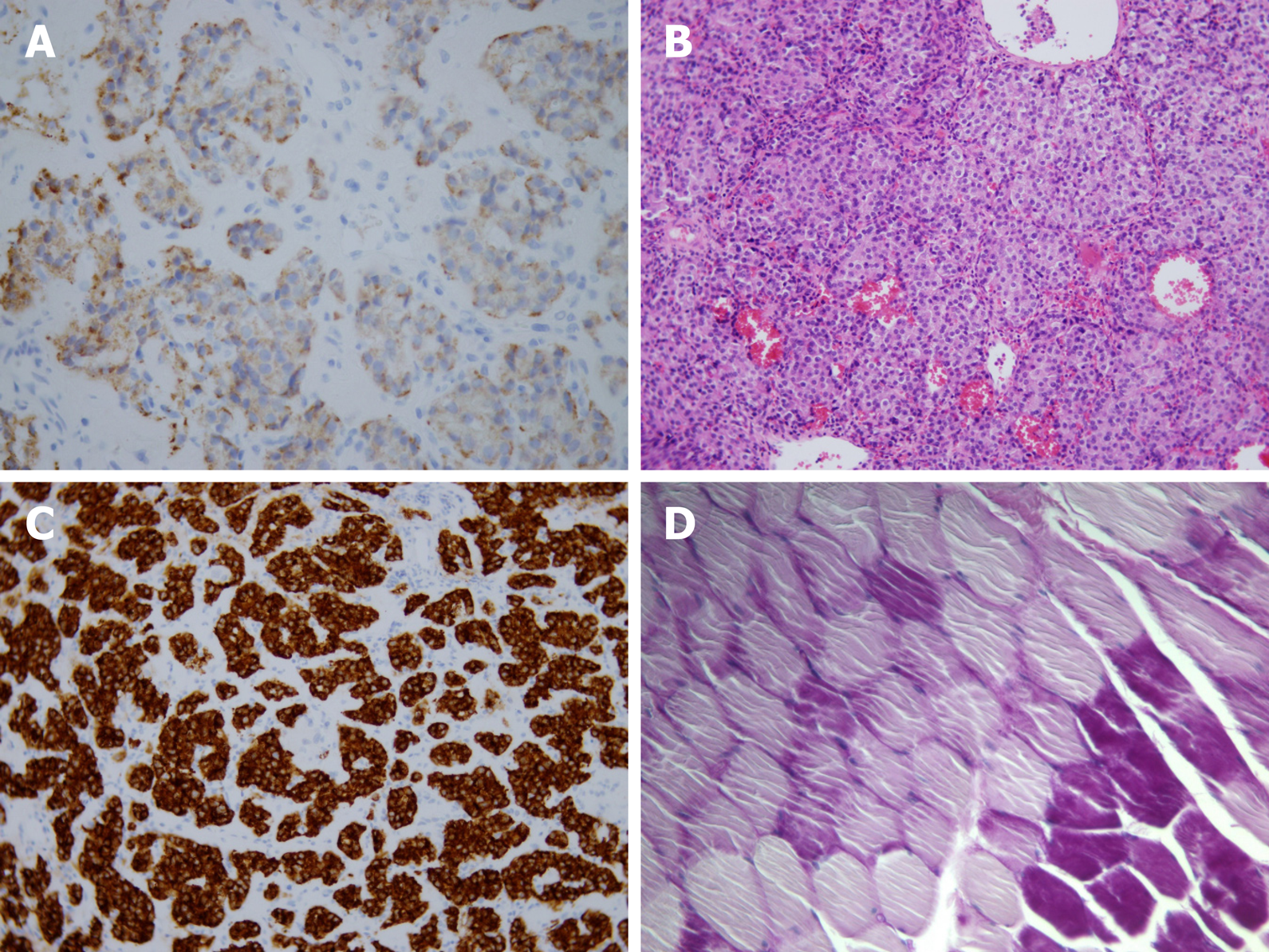
Figure 3 Histological and immunohistochemical findings of the insulinoma and histological findings of muscle biopsy.
A: Positive insulin immunostaining; B: Hematoxylin and eosin staining; C: Positive synaptophysin immunostaining; D: Glycogen deficit (light violet areas) in muscle syncytia (musculus quadriceps femoris), PAS stain, 20 ×.
- Citation: Prídavková D, Samoš M, Kyčina R, Adamicová K, Kalman M, Belicová M, Mokáň M. Insulinoma presenting with postprandial hypoglycemia and a low body mass index: A case report. World J Clin Cases 2020; 8(18): 4169-4176
- URL: https://www.wjgnet.com/2307-8960/full/v8/i18/4169.htm
- DOI: https://dx.doi.org/10.12998/wjcc.v8.i18.4169


