©The Author(s) 2020.
World J Clin Cases. Sep 26, 2020; 8(18): 4162-4168
Published online Sep 26, 2020. doi: 10.12998/wjcc.v8.i18.4162
Published online Sep 26, 2020. doi: 10.12998/wjcc.v8.i18.4162
Figure 1 Upper airway mucosa lesions.
A: Axial computed tomography (CT) (lung window) showing circumferential irregular thickening of the tracheal wall - subglottic region; B: Axial CT (lung window) demonstrating the presence of a tracheal mucosa nodule-carina level.
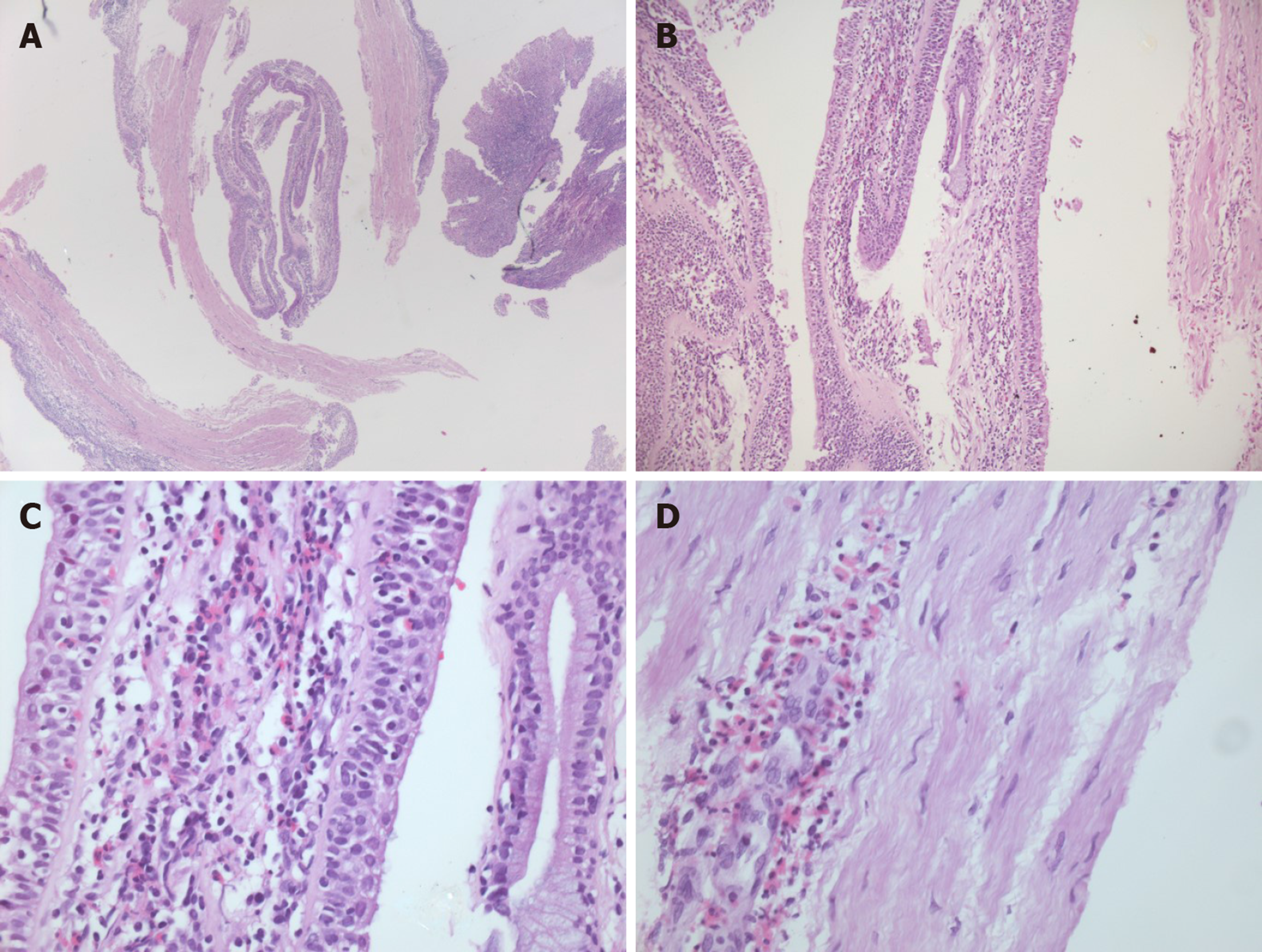
Figure 2 Hematoxylin-eosin stained tracheal mucosal biopsies demonstrating the presence of eosinophil-rich infiltrate.
A: Magnification 25 ×; B: Magnification 100 ×; C: Magnification 200 ×; D: Magnification 400 ×.
- Citation: Cernomaz AT, Bordeianu G, Terinte C, Gavrilescu CM. Nonasthmatic eosinophilic bronchitis in an ulcerative colitis patient – a putative adverse reaction to mesalazine: A case report and review of literature. World J Clin Cases 2020; 8(18): 4162-4168
- URL: https://www.wjgnet.com/2307-8960/full/v8/i18/4162.htm
- DOI: https://dx.doi.org/10.12998/wjcc.v8.i18.4162


