©The Author(s) 2020.
World J Clin Cases. Sep 26, 2020; 8(18): 4151-4161
Published online Sep 26, 2020. doi: 10.12998/wjcc.v8.i18.4151
Published online Sep 26, 2020. doi: 10.12998/wjcc.v8.i18.4151
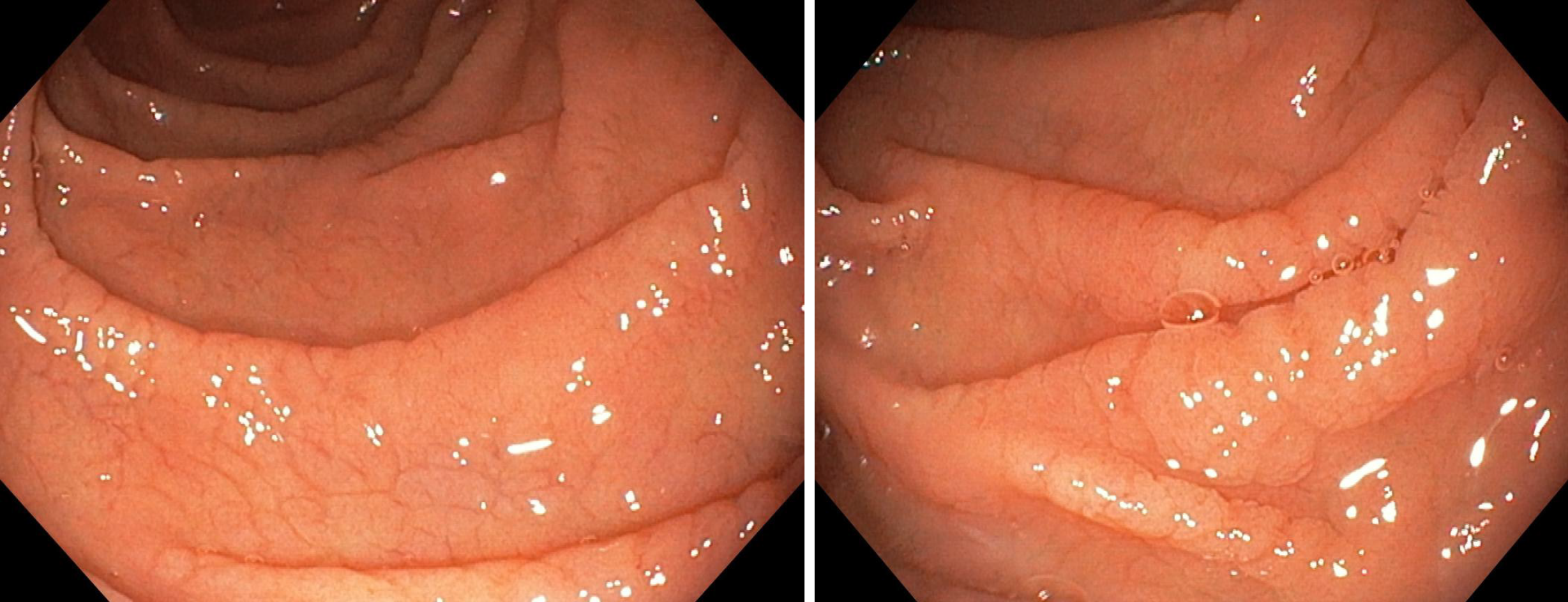
Figure 1 Endoscopic images showing mucosal fissures and scalloping in the distal duodenum.
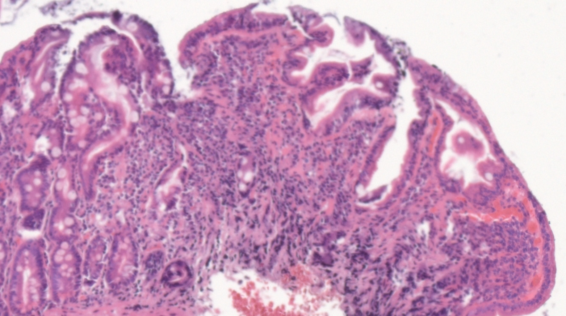
Figure 2 Hematoxilin-eosin stain of duodenal biopsy sample showing marked villous atrophy with crypt hyperplasia and intraepithelial lymphocytosis, corresponding to Marsh 3c classification.
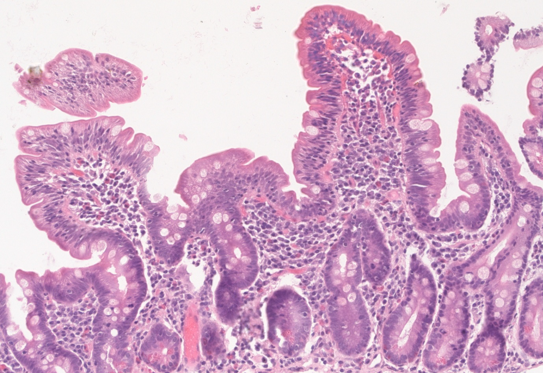
Figure 3 Hematoxilin-eosin stain of follow-up biopsy revealing restoring of normal villous architecture but with persisting intraepithelial lymphocytosis.
- Citation: Balaban DV, Mihai A, Dima A, Popp A, Jinga M, Jurcut C. Celiac disease and Sjögren’s syndrome: A case report and review of literature. World J Clin Cases 2020; 8(18): 4151-4161
- URL: https://www.wjgnet.com/2307-8960/full/v8/i18/4151.htm
- DOI: https://dx.doi.org/10.12998/wjcc.v8.i18.4151


