©The Author(s) 2020.
World J Clin Cases. Sep 6, 2020; 8(17): 3828-3834
Published online Sep 6, 2020. doi: 10.12998/wjcc.v8.i17.3828
Published online Sep 6, 2020. doi: 10.12998/wjcc.v8.i17.3828
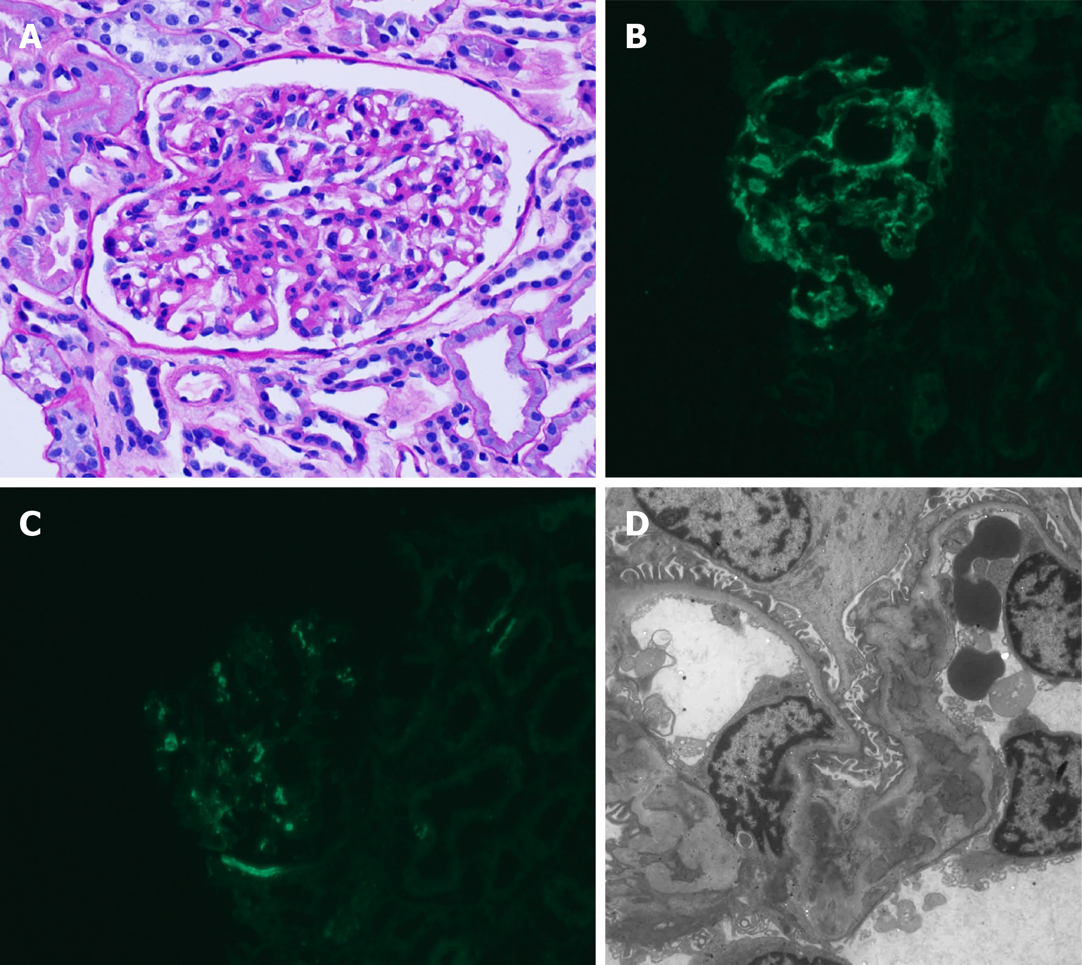
Figure 1 Kidney histopathology.
A: Light microscopy, mesangial hypercellularity in a glomerulus; B: Immunofluorescence microscopy, the staining of immunoglobulin A (3+); C: Immunofluorescence microscopy; the staining of C3 (1+); D: Electron microscopy, mesangial electron- dense deposits.
Figure 2 Liver histopathology.
A: Hematoxylin and eosin staining (magnification, 40 ×), submassive necrosis of the liver parenchyma with prominent bile duct proliferation; B: Hematoxylin and eosin staining (magnification, 100 ×), canalicular type cholestasis.
- Citation: Jeon YH, Kim DW, Lee SJ, Park YJ, Kim HJ, Han M, Kim IY, Lee DW, Song SH, Lee SB, Seong EY. Autoimmune hepatitis in a patient with immunoglobulin A nephropathy: A case report. World J Clin Cases 2020; 8(17): 3828-3834
- URL: https://www.wjgnet.com/2307-8960/full/v8/i17/3828.htm
- DOI: https://dx.doi.org/10.12998/wjcc.v8.i17.3828


