©The Author(s) 2020.
World J Clin Cases. Aug 26, 2020; 8(16): 3548-3552
Published online Aug 26, 2020. doi: 10.12998/wjcc.v8.i16.3548
Published online Aug 26, 2020. doi: 10.12998/wjcc.v8.i16.3548
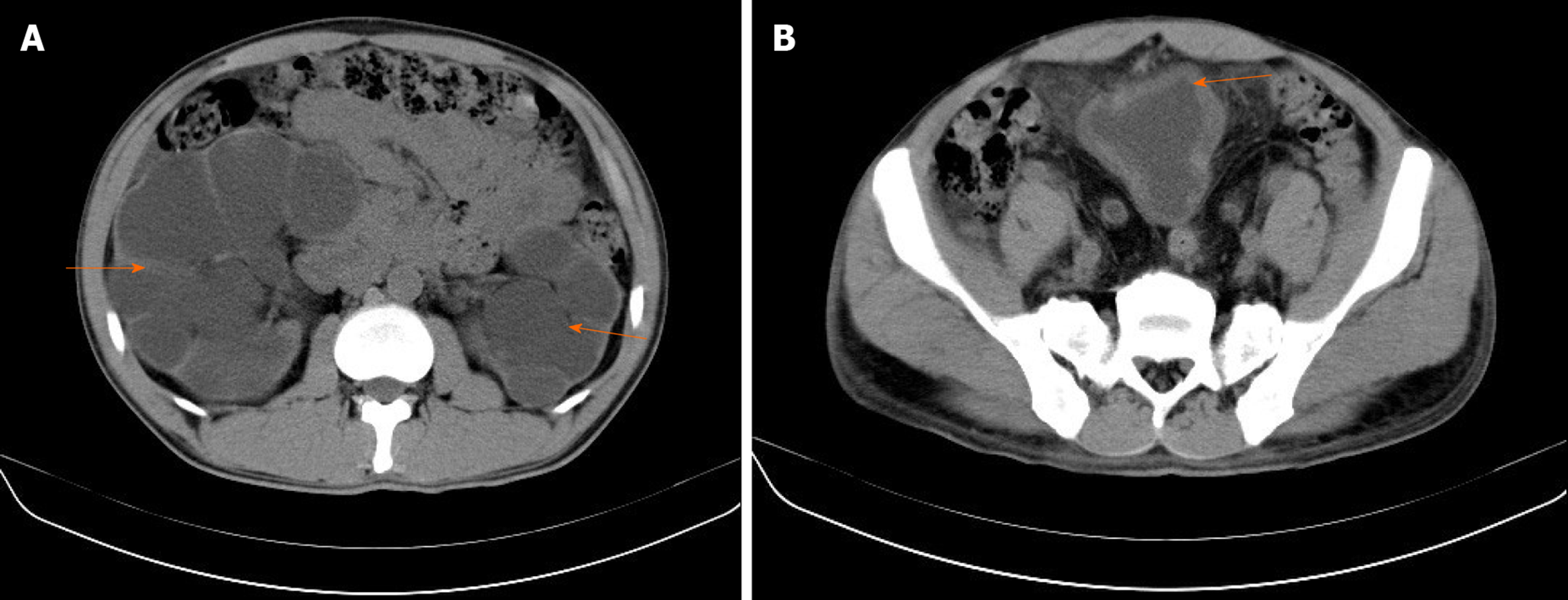
Figure 1 Computed tomography examination.
A: Computed tomography showed bilateral hydronephrosis (orange arrow); B: A thick walled urinary bladder (orange arrow) with increased fat density around the bladder.
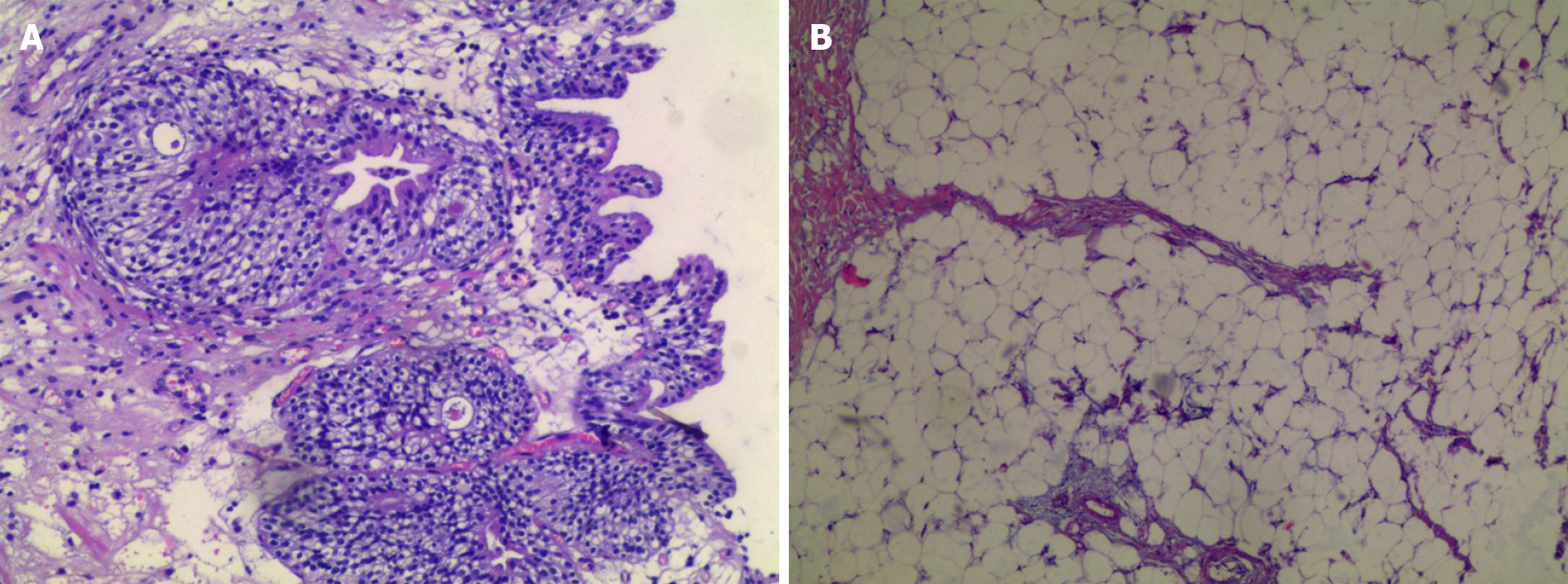
Figure 2 Histopathological examination.
A: Tissue biopsy of the bladder demonstrated cystitis glandularis; B: Histopathological examination of the fat showed a benign lipomatous lesion.
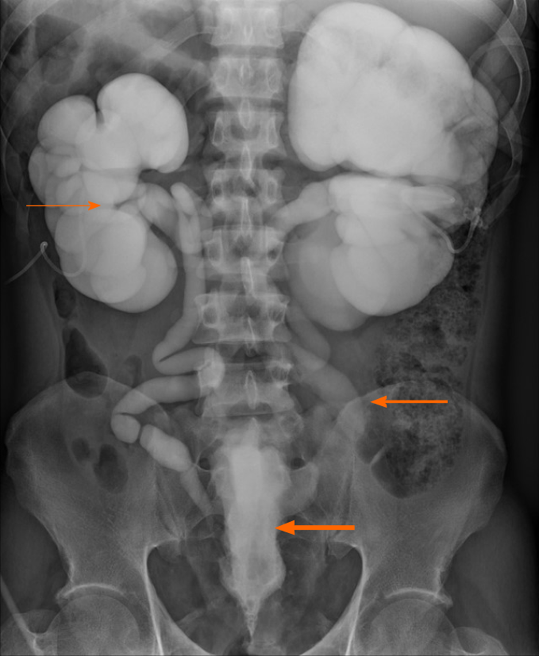
Figure 3 Anterograde urography revealed bilateral hydronephrosis (thin orange arrow) and tortuous dilated ureter (orange arrow), symmetrical compression and elevation of the bladder (a pear-shaped bladder) (thick orange arrow).
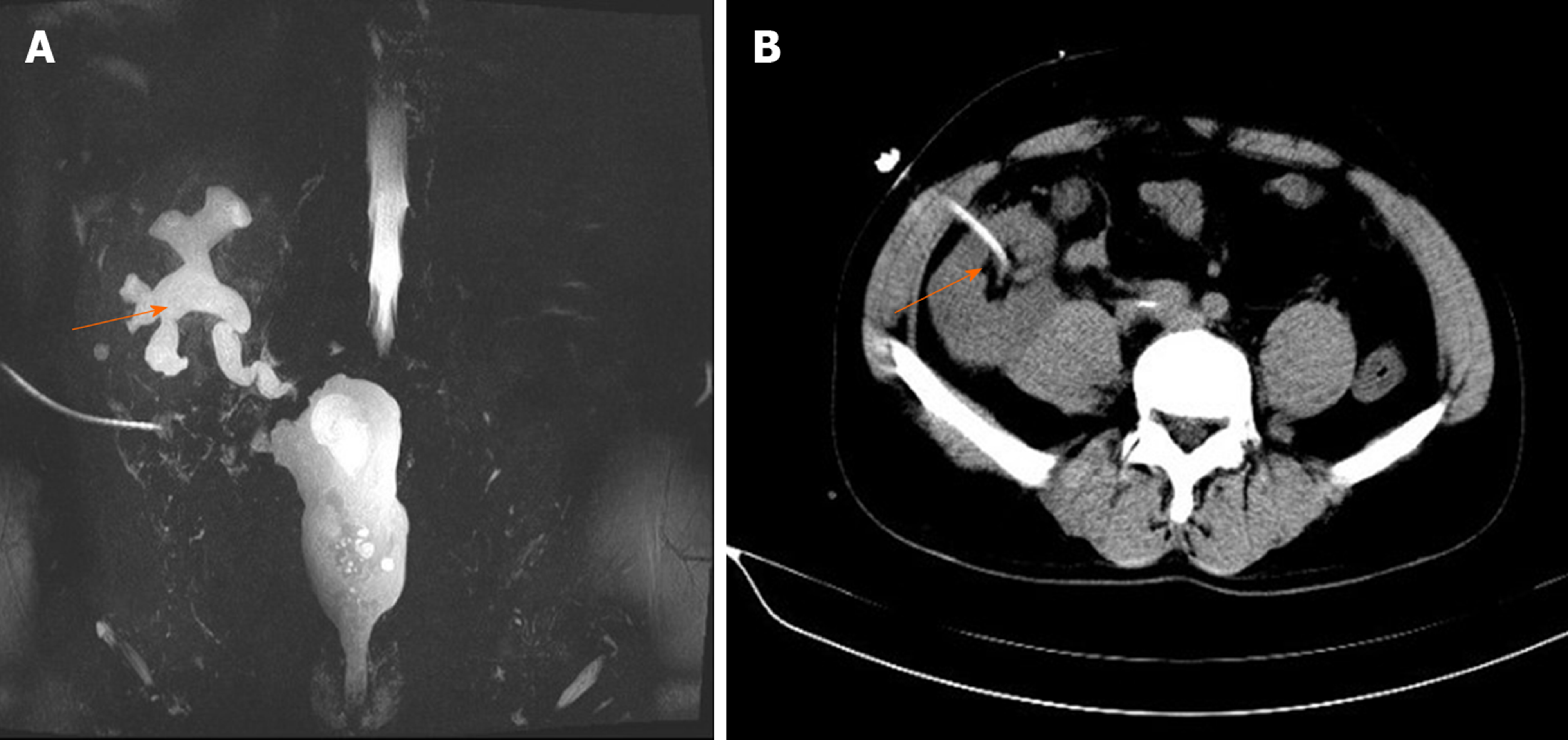
Figure 4 Magnetic resonance urography.
A: Magnetic resonance urography show mild hydronephrosis in the graft kidney at 6 mo postoperation (orange arrow); B: Graft percutaneous nephrostomy was performed under ultrasonographic guidance for urinary diversion (orange arrow).
- Citation: Zhao J, Fu YX, Feng G, Mo CB. Pelvic lipomatosis and renal transplantation: A case report. World J Clin Cases 2020; 8(16): 3548-3552
- URL: https://www.wjgnet.com/2307-8960/full/v8/i16/3548.htm
- DOI: https://dx.doi.org/10.12998/wjcc.v8.i16.3548


