Copyright
©The Author(s) 2020.
World J Clin Cases. Aug 26, 2020; 8(16): 3527-3533
Published online Aug 26, 2020. doi: 10.12998/wjcc.v8.i16.3527
Published online Aug 26, 2020. doi: 10.12998/wjcc.v8.i16.3527
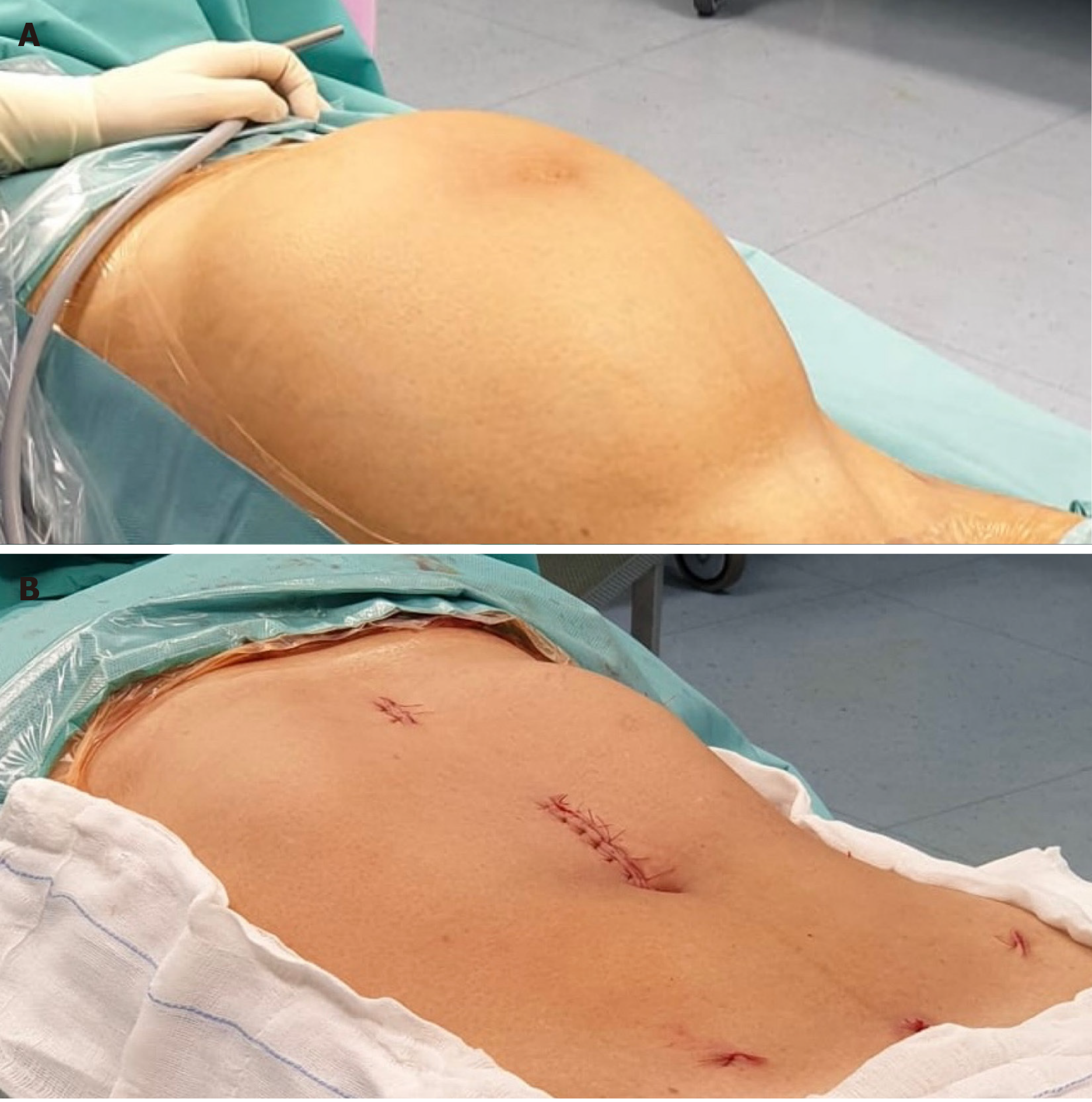
Figure 1 View of the abdomen before and after surgery.
A: Enlarged abdomen with the patient lying supine before surgery; B: View of the abdomen after surgery.
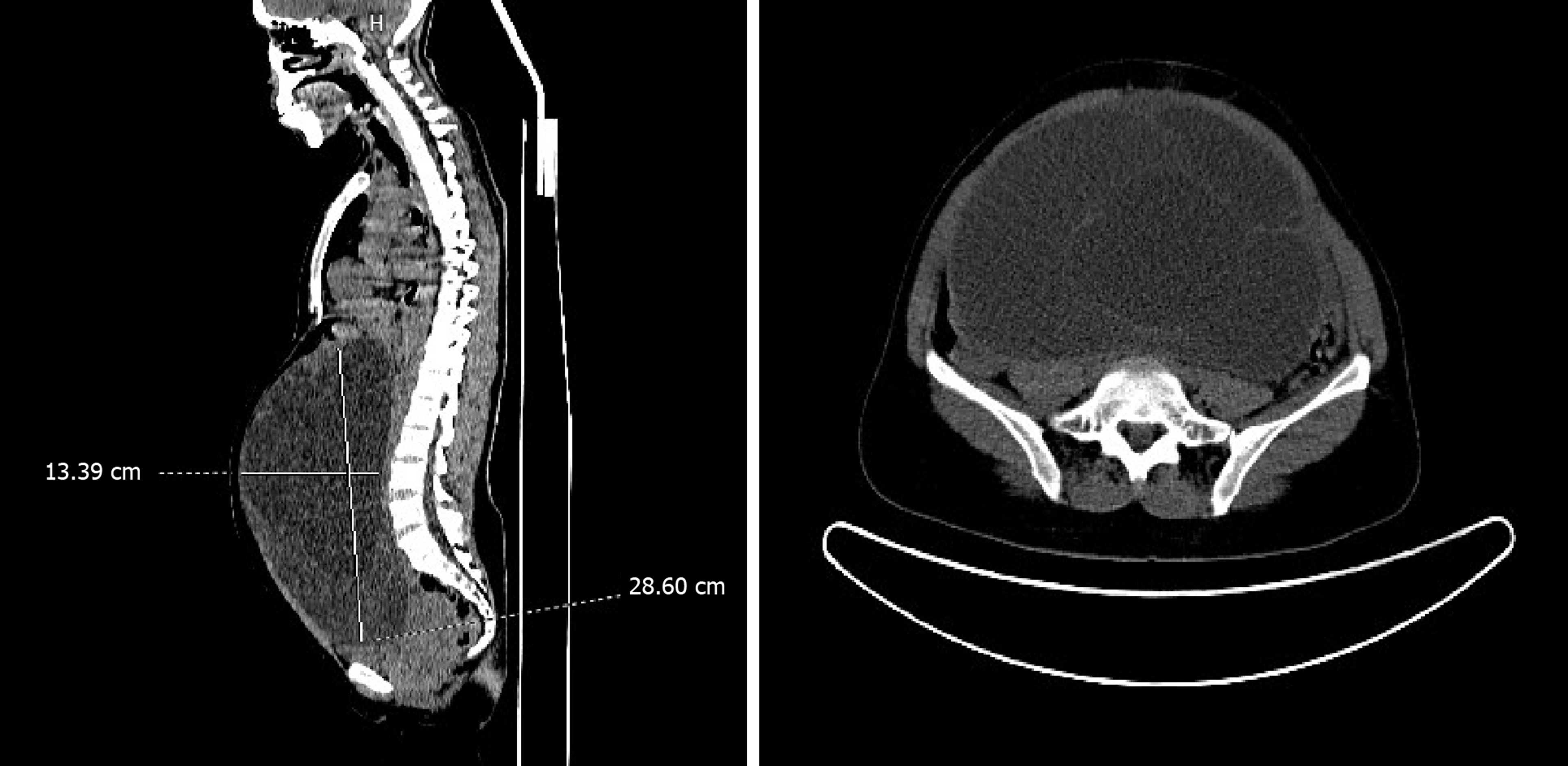
Figure 2 Preoperative computed tomography imaging scan.
Computed tomography images showing the sagittal and transverse view of the multi-locular giant cyst.
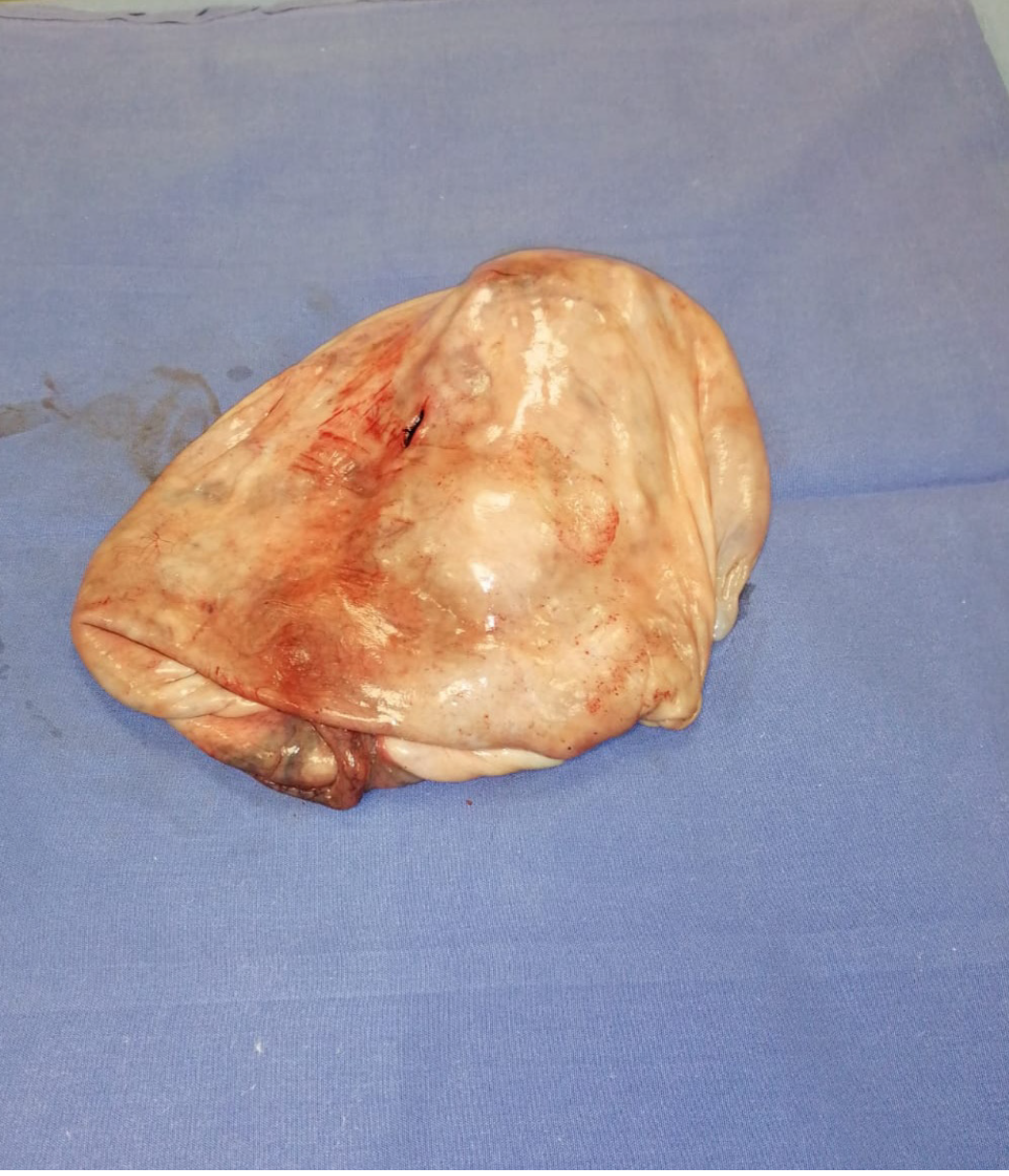
Figure 3 The cyst wall after extraction from the abdomen.
- Citation: Sanna E, Madeddu C, Melis L, Nemolato S, Macciò A. Laparoscopic management of a giant mucinous benign ovarian mass weighing 10150 grams: A case report. World J Clin Cases 2020; 8(16): 3527-3533
- URL: https://www.wjgnet.com/2307-8960/full/v8/i16/3527.htm
- DOI: https://dx.doi.org/10.12998/wjcc.v8.i16.3527


