©The Author(s) 2020.
World J Clin Cases. Jun 26, 2020; 8(12): 2667-2673
Published online Jun 26, 2020. doi: 10.12998/wjcc.v8.i12.2667
Published online Jun 26, 2020. doi: 10.12998/wjcc.v8.i12.2667
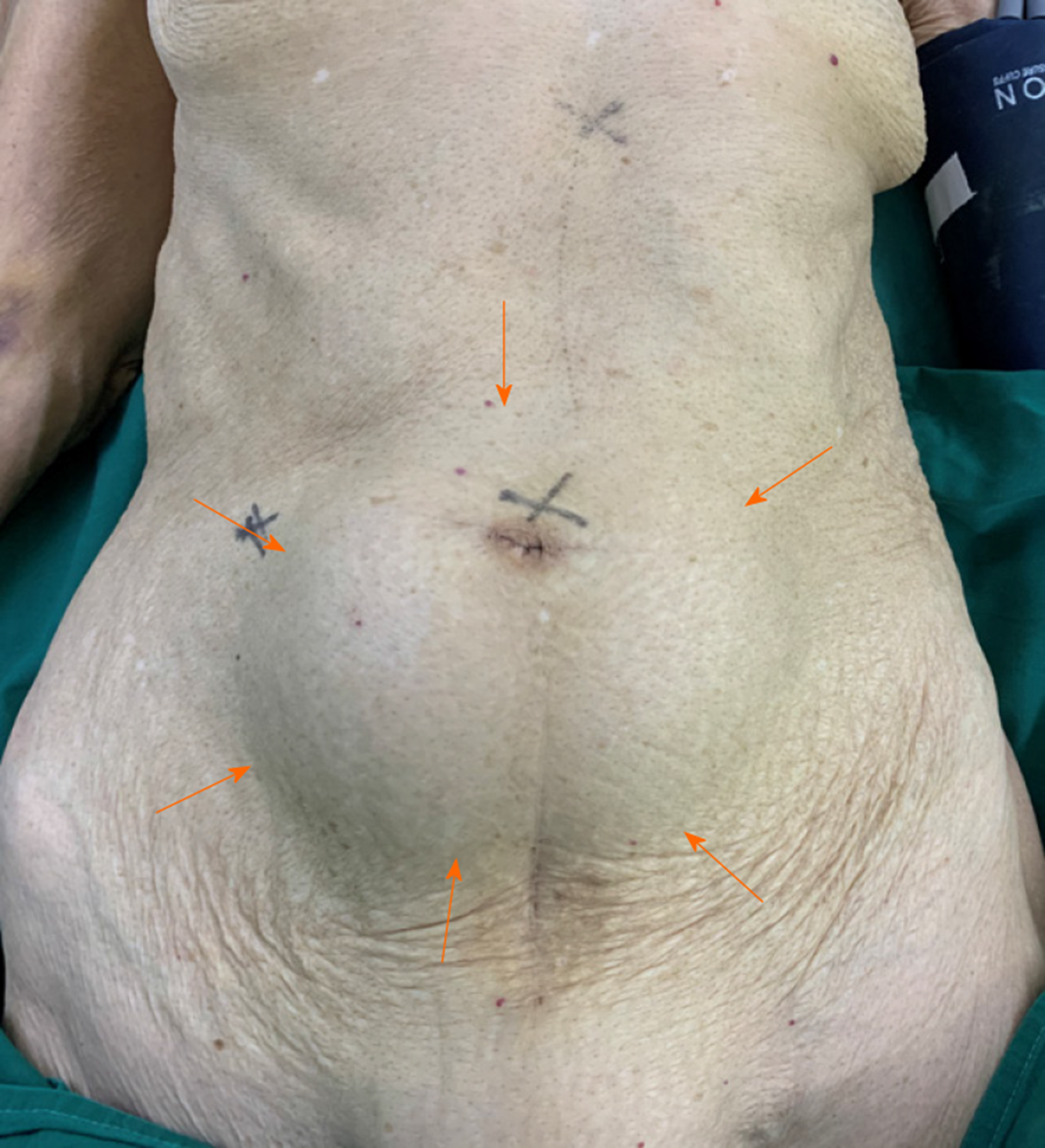
Figure 1 Physical examination showing a mass in the region around the umbilicus.
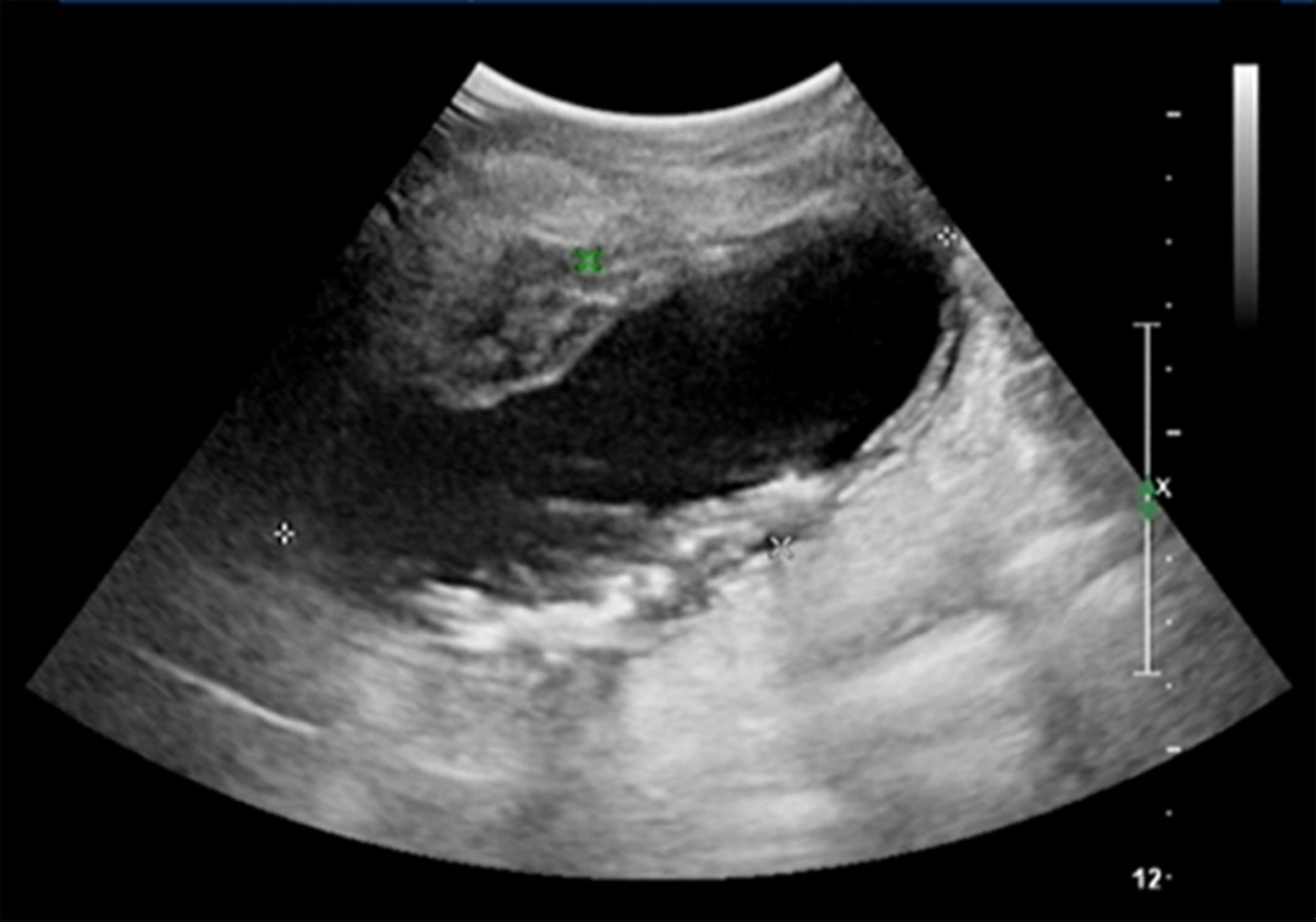
Figure 2 Ultrasonography showing a cystic mass in the lower abdomen and upper pelvis with multiple highly echoic nodules.
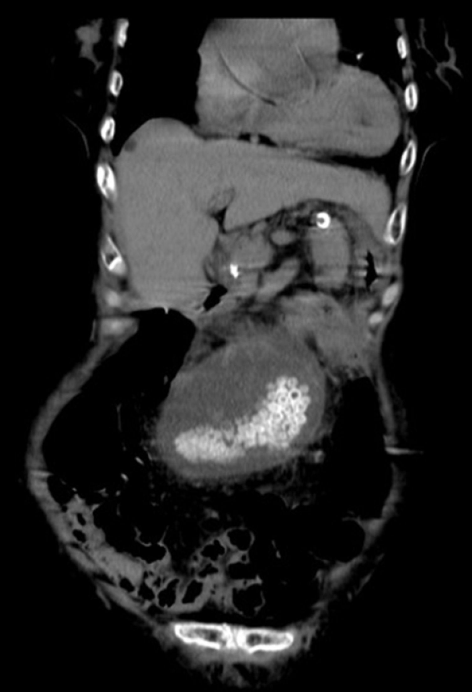
Figure 3 Computed tomography showing a cystic mass in the lower abdomen and upper pelvis with multiple nodules of high-density calcifications.
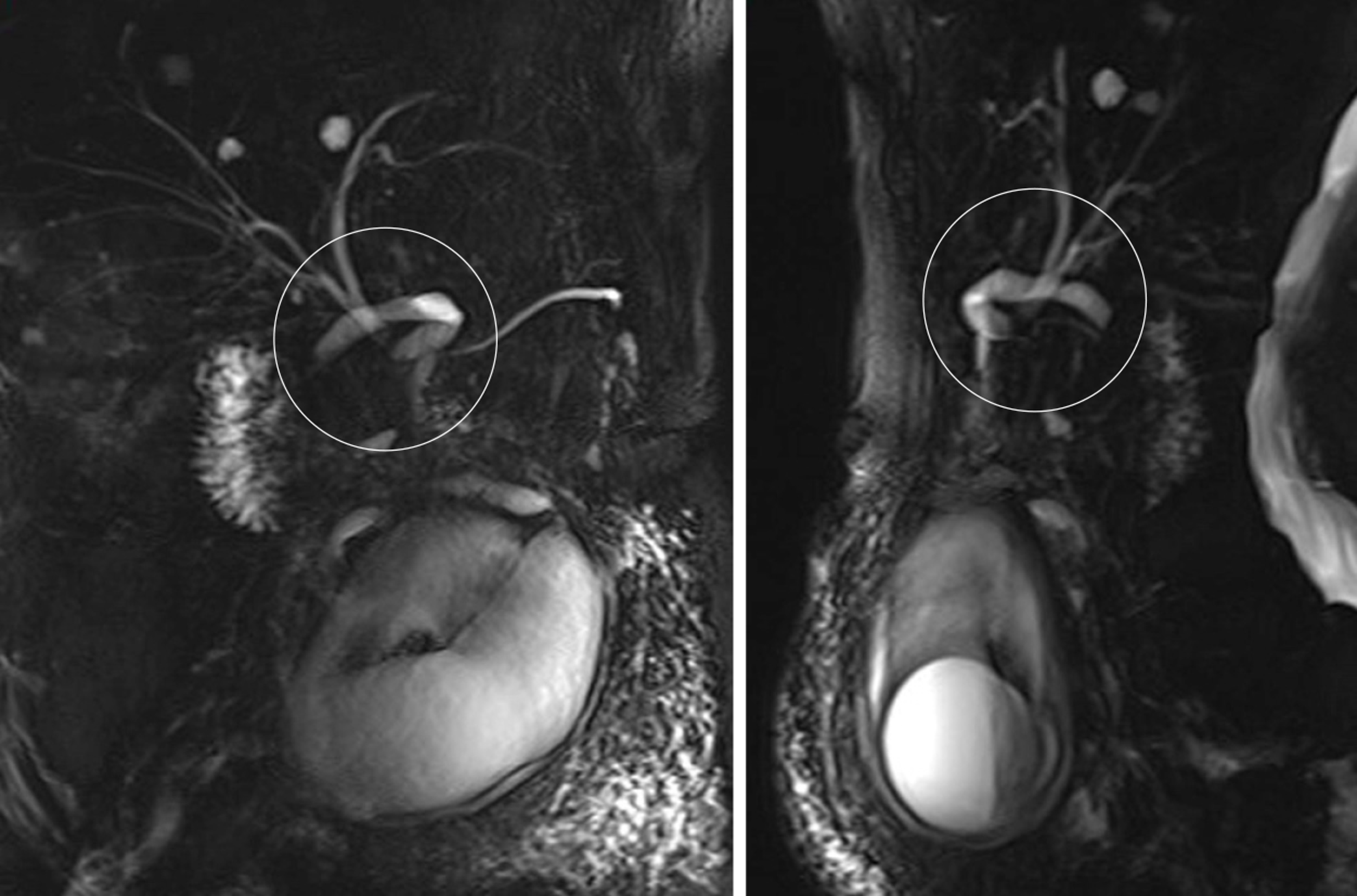
Figure 4 Magnetic resonance cholangiopancreatography showing a V-shaped distortion of the extrahepatic bile ducts and a particularly extended twisted cystic duct, called “twisting signs”.
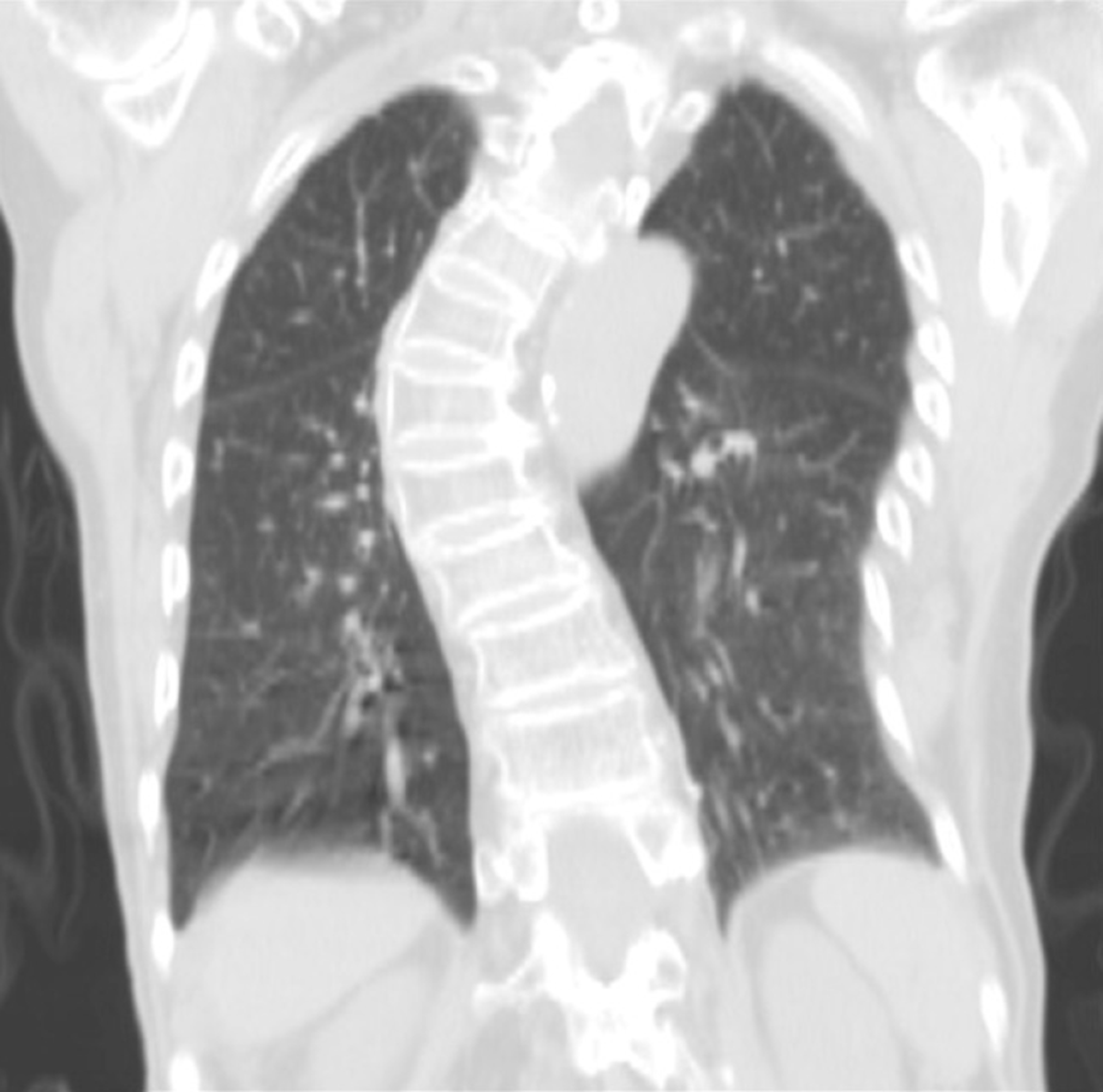
Figure 5 Chest computed tomography before surgery showing spinal deformities.
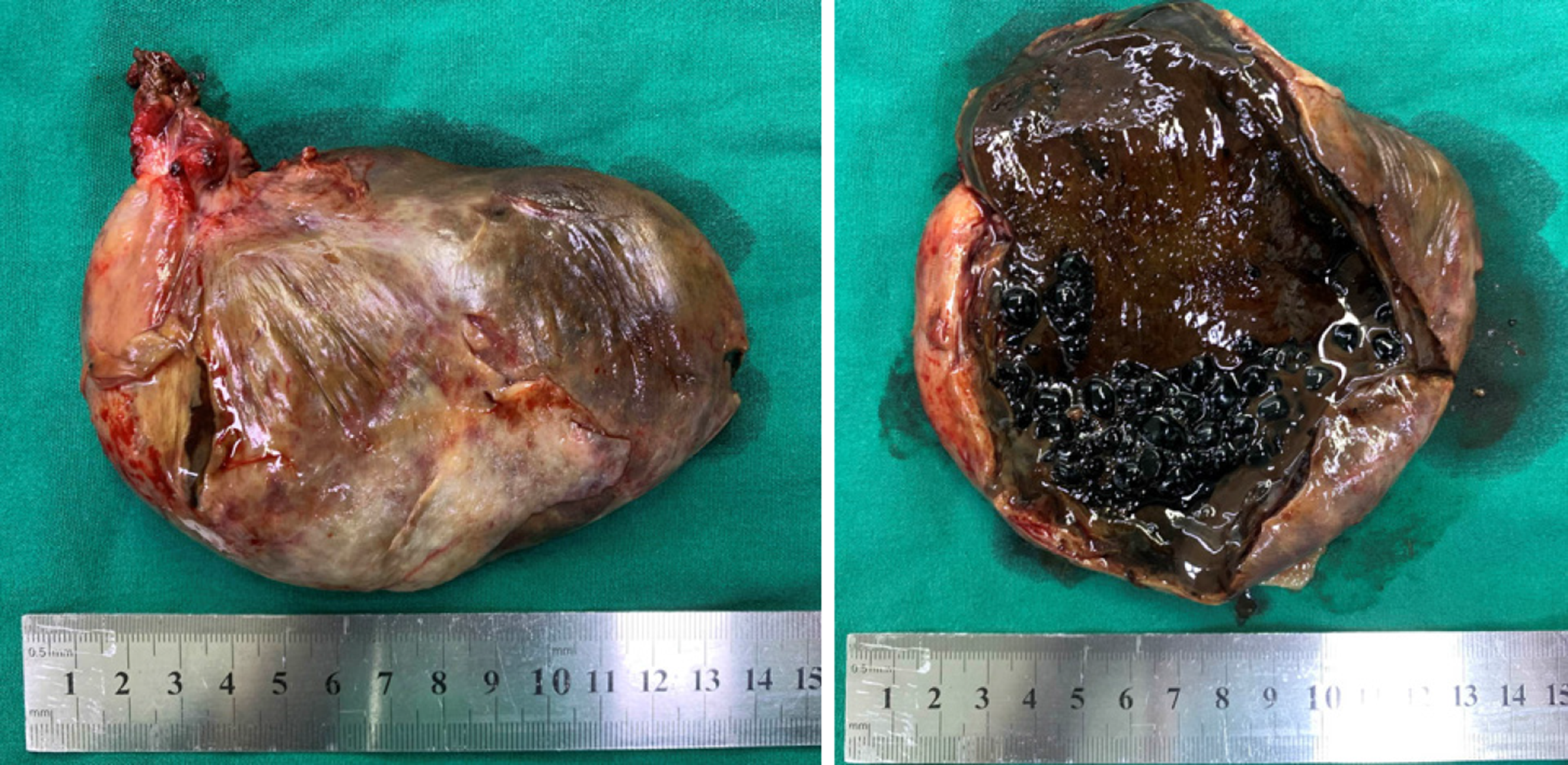
Figure 6 Decompressed gallbladder.
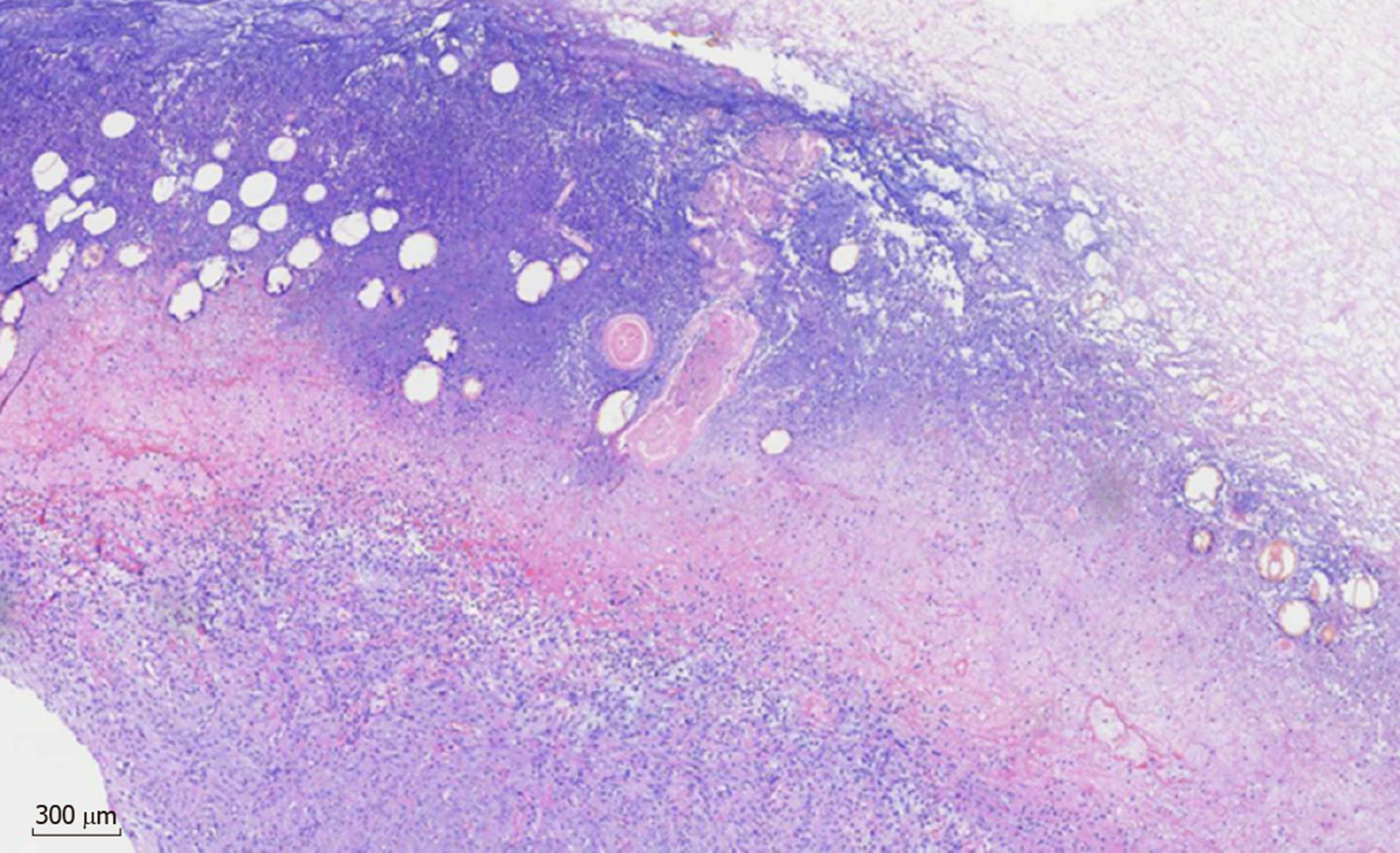
Figure 7 Histopathological findings showing acute gangrenous cholecystitis.
- Citation: Chai JS, Wang X, Li XZ, Yao P, Yan ZZ, Zhang HJ, Ning JY, Cao YB. Presentation of gallbladder torsion at an abnormal position: A case report. World J Clin Cases 2020; 8(12): 2667-2673
- URL: https://www.wjgnet.com/2307-8960/full/v8/i12/2667.htm
- DOI: https://dx.doi.org/10.12998/wjcc.v8.i12.2667


