©The Author(s) 2020.
World J Clin Cases. Jun 26, 2020; 8(12): 2641-2646
Published online Jun 26, 2020. doi: 10.12998/wjcc.v8.i12.2641
Published online Jun 26, 2020. doi: 10.12998/wjcc.v8.i12.2641
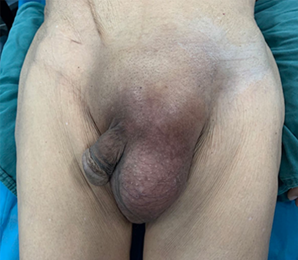
Figure 1 Physical examination.
A left scrotal mass measuring 15 cm was noted.
Figure 2 Intraoperative finding.
A and B: Coronal (A) and sagittal (B) views of contrast-enhanced magnetic resonance imaging showing a hydrocele testis and a heterogeneously enhanced lesion with a relatively well-defined margin; C: Intraoperative finding of the spermatic cord tumor and a secondary hydrocele testis.
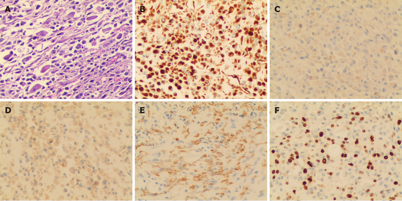
Figure 3 Histopathological and immunohistochemical analyses.
A: Formalin-fixed paraffin-embedded tumor tissue was stained with hematoxylin and eosin (100 ×); B: Vimentin (200 ×, cytoplasmic and membranous staining); C: Desmin (200 ×, cytoplasmic staining); D: Myogenin (200 ×, nuclear staining); E: Smooth muscle actin (200 ×, cytoplasmic and membranous staining); F: Ki-67 (200 ×, approximately 40% positivity).
- Citation: Chen X, Zou C, Yang C, Gao L, Bi LK, Xie DD, Yu DX. Pleomorphic rhabdomyosarcoma of the spermatic cord and a secondary hydrocele testis: A case report. World J Clin Cases 2020; 8(12): 2641-2646
- URL: https://www.wjgnet.com/2307-8960/full/v8/i12/2641.htm
- DOI: https://dx.doi.org/10.12998/wjcc.v8.i12.2641


