©The Author(s) 2020.
World J Clin Cases. Jun 6, 2020; 8(11): 2392-2398
Published online Jun 6, 2020. doi: 10.12998/wjcc.v8.i11.2392
Published online Jun 6, 2020. doi: 10.12998/wjcc.v8.i11.2392
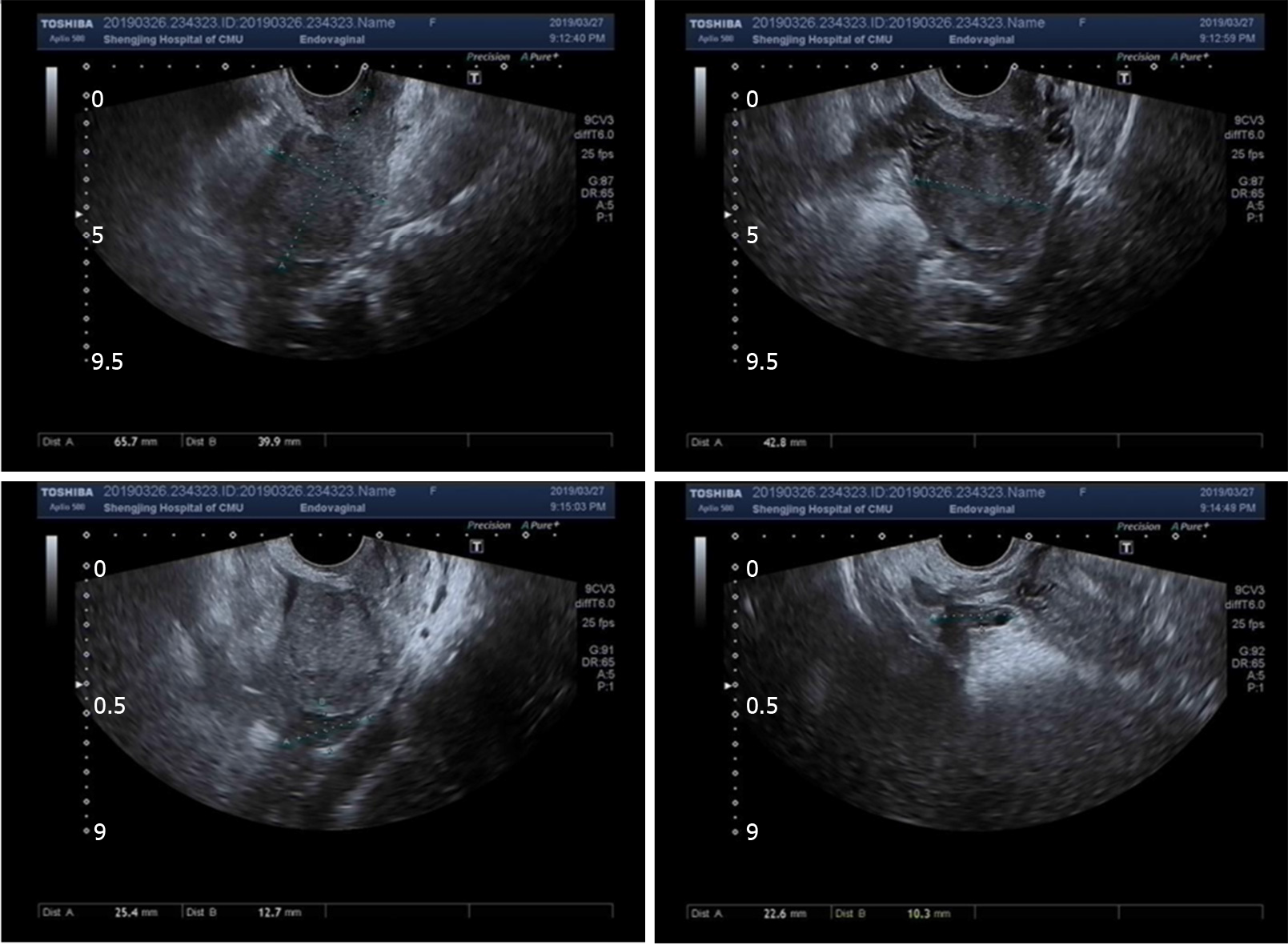
Figure 1 Transvaginal ultrasound showed an endometrium approximately 0.
7 mm thick with slightly uneven echo and no other abnormalities.
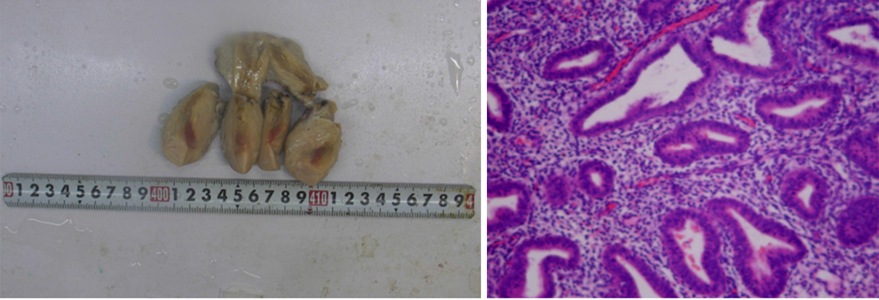
Figure 2 The paraffin pathology of the uterus.
A: Uterus; B: Postoperative pathological examination showed proliferative endometrial changes with multiple focal lymphocyte infiltrations in the interstitium (100 ×); some lymphocytes were mixed with plasma cells. In the red area of the uterine isthmus, the interstitial small blood vessels dilated, and the wall thickened like hyaline degeneration. The pathological diagnosis was local fibrous hyperplasia and inflammatory changes in the uterine isthmus. The blood vessels in the superficial myometrium of the uterus were widely transparent and sclerotic.
- Citation: Zhang Y, Ma NY, Pang XA. Uterine incision dehiscence 3 mo after cesarean section causing massive bleeding: A case report. World J Clin Cases 2020; 8(11): 2392-2398
- URL: https://www.wjgnet.com/2307-8960/full/v8/i11/2392.htm
- DOI: https://dx.doi.org/10.12998/wjcc.v8.i11.2392


