©The Author(s) 2020.
World J Clin Cases. Jun 6, 2020; 8(11): 2380-2386
Published online Jun 6, 2020. doi: 10.12998/wjcc.v8.i11.2380
Published online Jun 6, 2020. doi: 10.12998/wjcc.v8.i11.2380
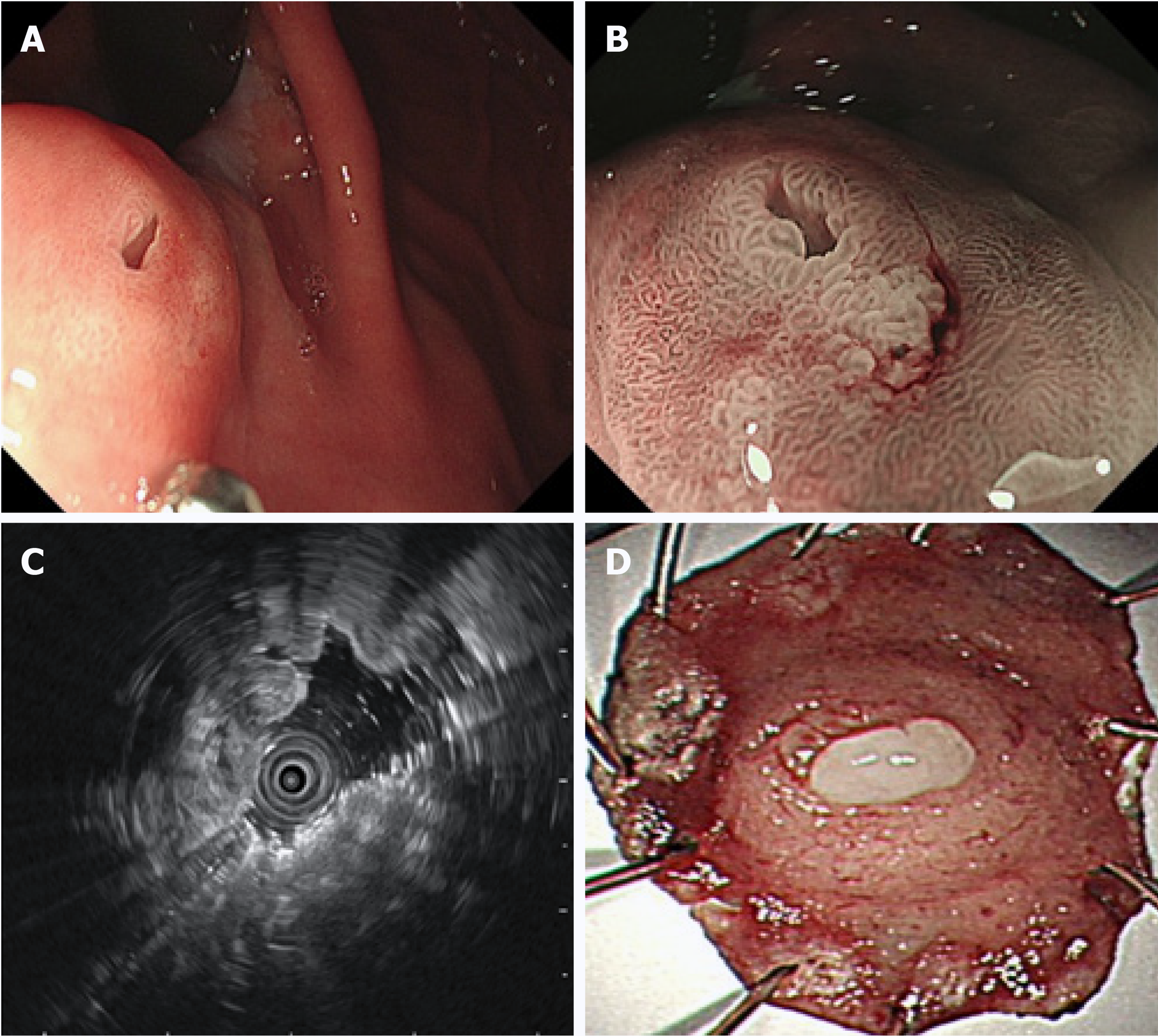
Figure 1 Endoscopic findings.
A: A 10 mm submucosal tumor-like elevated lesion with an opening in the posterior wall of the upper part of the gastric body was observed by white light endoscopy; B: A regular microvascular pattern was observed using magnifying endoscopy with narrow-band imaging (magnification: × 40); C: Isoechoic mass (10.6 mm × 5.5 mm) with multiple cysts could be observed in the submucosal layer with intact muscularis using endoscopic ultrasound; D: An elevated tumor measuring 13 mm × 10 mm with oozing white mucus could be observed in the endoscopic submucosal dissection specimen.
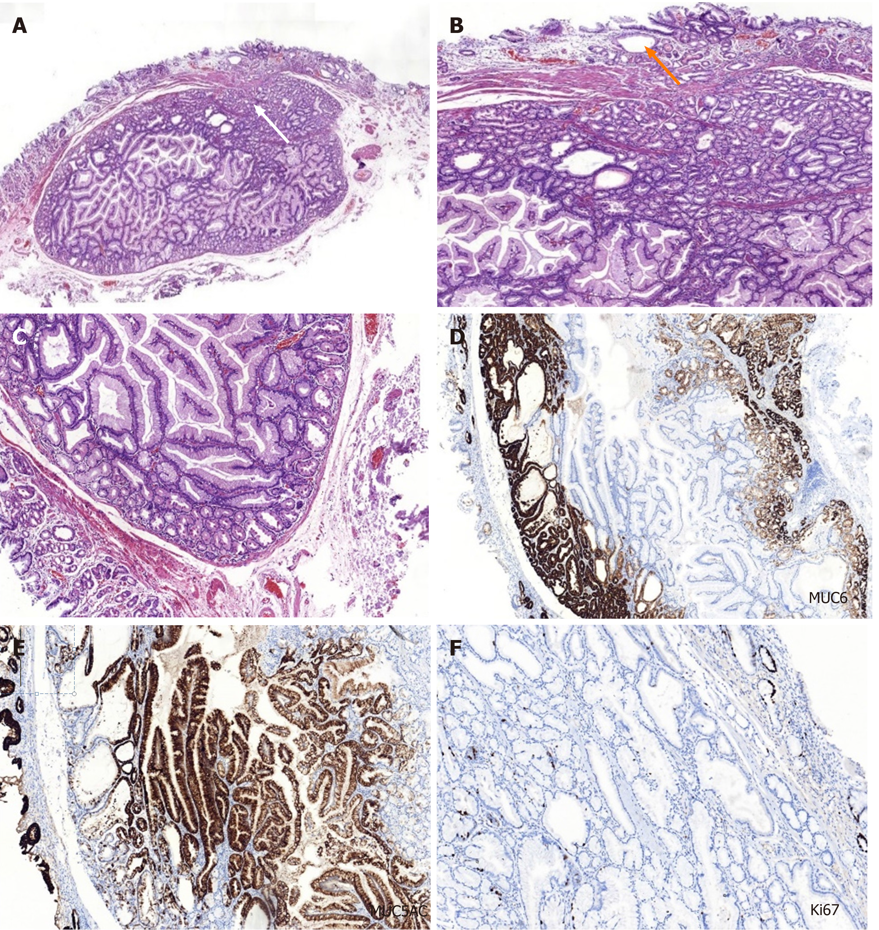
Figure 2 Histopathological and immunohistochemical findings of the pyloric gland adenoma.
A-C: Hematoxylin and eosin staining. The submucosal layer shows presence of nodular-like lesions with clear boundaries and increasing number of glands in the lamina propria of the orifice (white arrow) (A; magnification: × 20). Some glandular cavity was irregular, with cystic expansion (orange arrow) and interstitial edema (B; magnification: × 200). The cuboidal or low columnar epithelial cells line the mucus-rich tubular glands in these lesions (C; magnification: × 200); D-F: Immunohistochemical staining. The glandular epithelial cells revealed strongly positive for mucin mucin 6 (D) and mucin 5AC (E) and sparsely positive for Ki67 (F). Magnification: D-F: × 100. MUC5AC: Mucin 5AC; MUC 6: Mucin 6.
- Citation: Min CC, Wu J, Hou F, Mao T, Li XY, Ding XL, Liu H. Gastric pyloric gland adenoma resembling a submucosal tumor: A case report. World J Clin Cases 2020; 8(11): 2380-2386
- URL: https://www.wjgnet.com/2307-8960/full/v8/i11/2380.htm
- DOI: https://dx.doi.org/10.12998/wjcc.v8.i11.2380


