©The Author(s) 2020.
World J Clin Cases. Jun 6, 2020; 8(11): 2312-2317
Published online Jun 6, 2020. doi: 10.12998/wjcc.v8.i11.2312
Published online Jun 6, 2020. doi: 10.12998/wjcc.v8.i11.2312
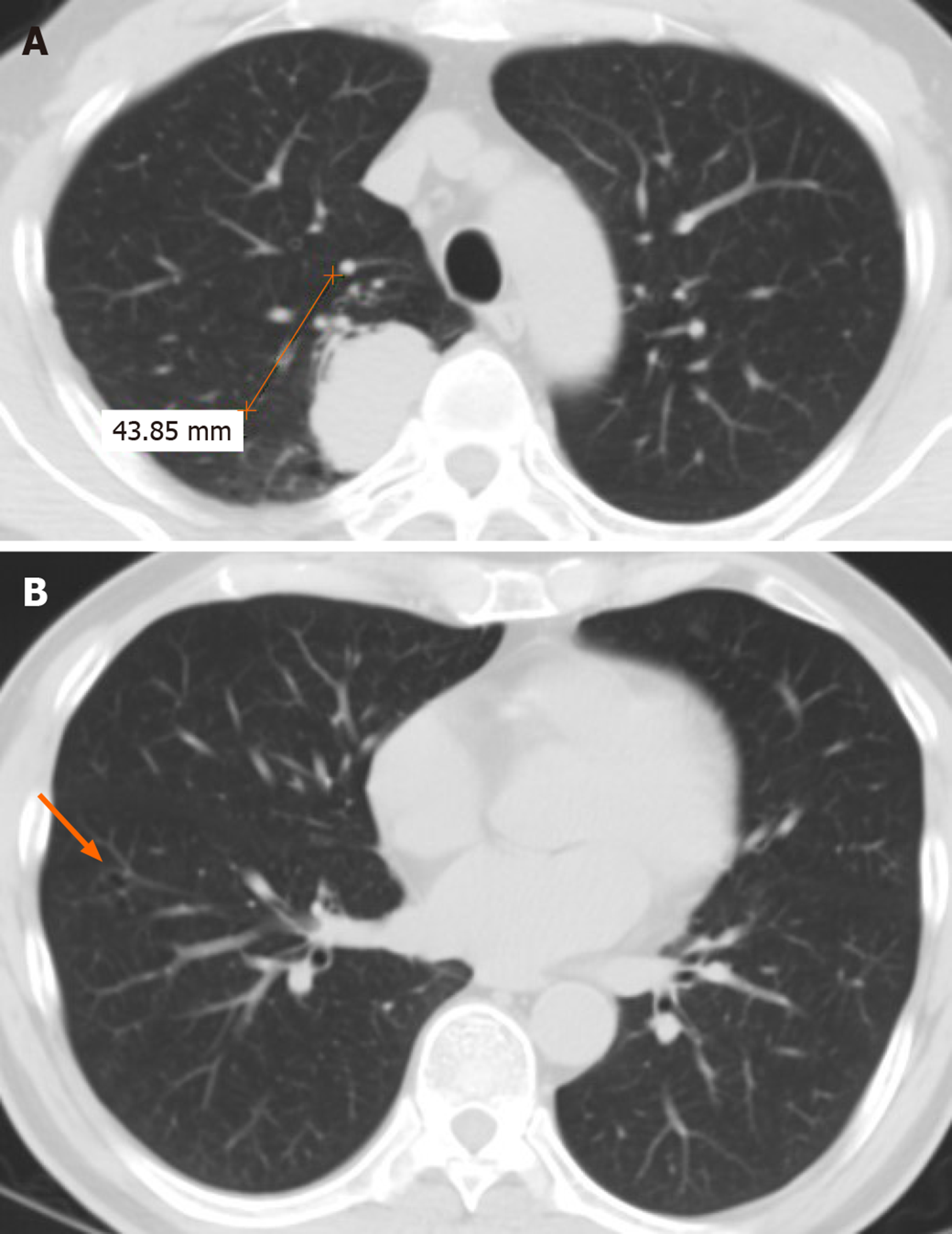
Figure 1 Chest computed tomography scan in 2007 showed a 43-mm solid lesion in the right upper lung and a small thin-walled cavity in the lower lobe.
A: A 43-mm solid lesion in the right upper lung; B: A small thin-walled cavity in the lower lobe (arrow).
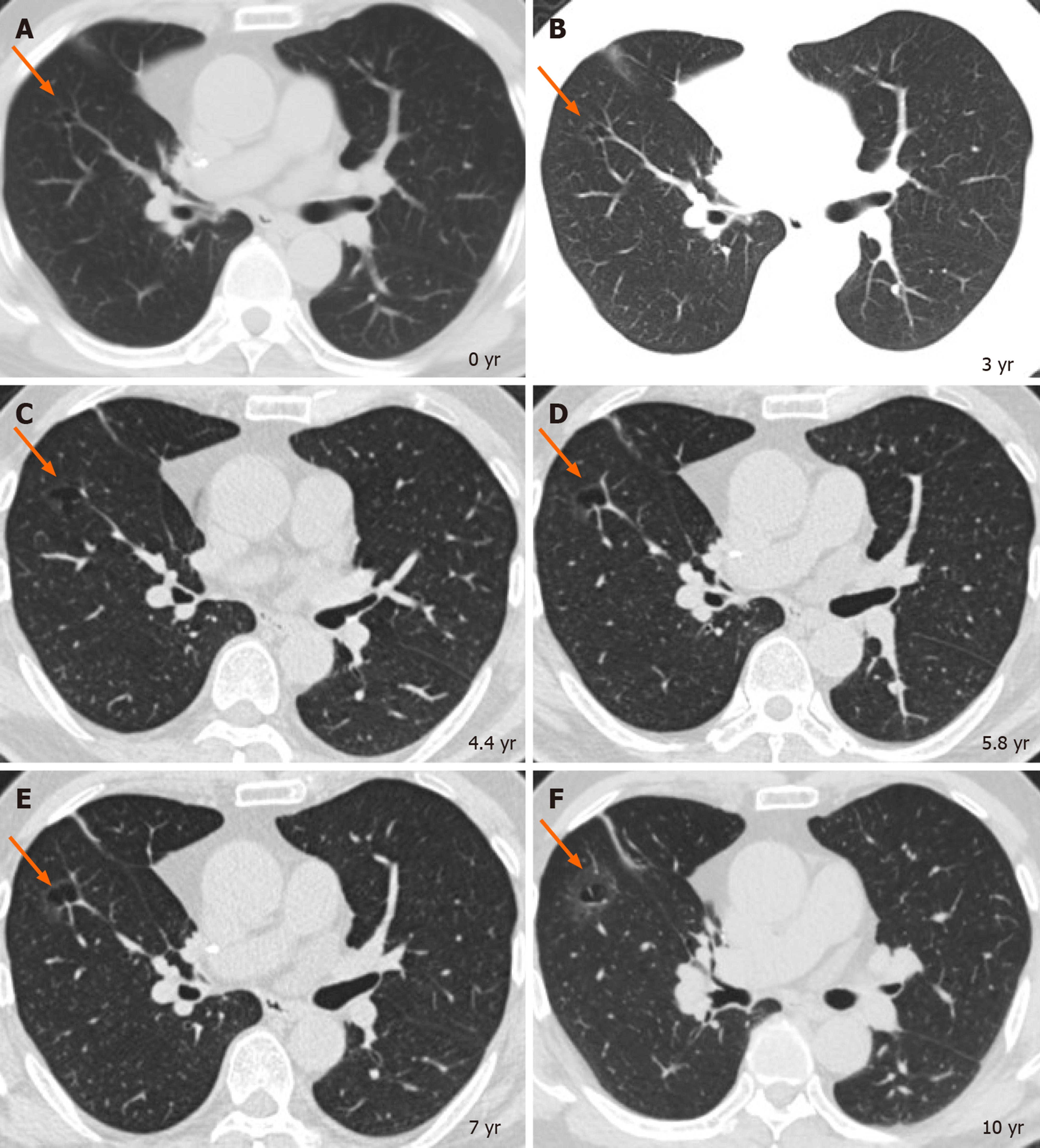
Figure 2 Serial computed tomography images over 10 years in this patientshowed the gradual evolution of an 11-mm right lower lobe thin-walled cystic airspace (A, arrow) into a 31-mm cystic airspace with a thick wall demonstrating as a GGO (F, arrow).
A: Before the first operation (2007); B: Three years later; C: 4.4 years later; D: 5.8 years later; E: 7 years later; F: 10 years later.
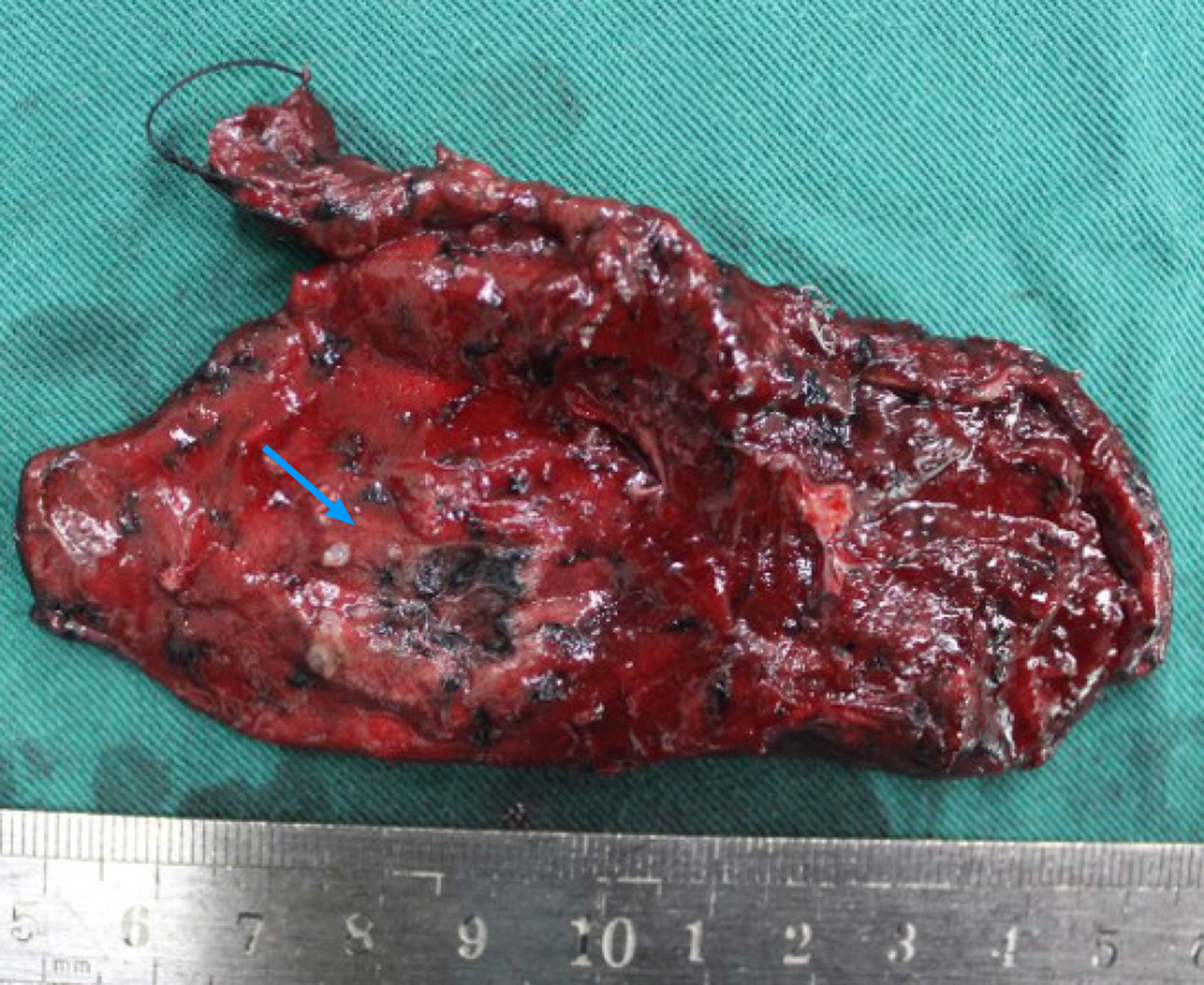
Figure 3 Surgical specimen showing a thick-walled cavity (arrow) with a malignant appearance in the surrounding tissue.
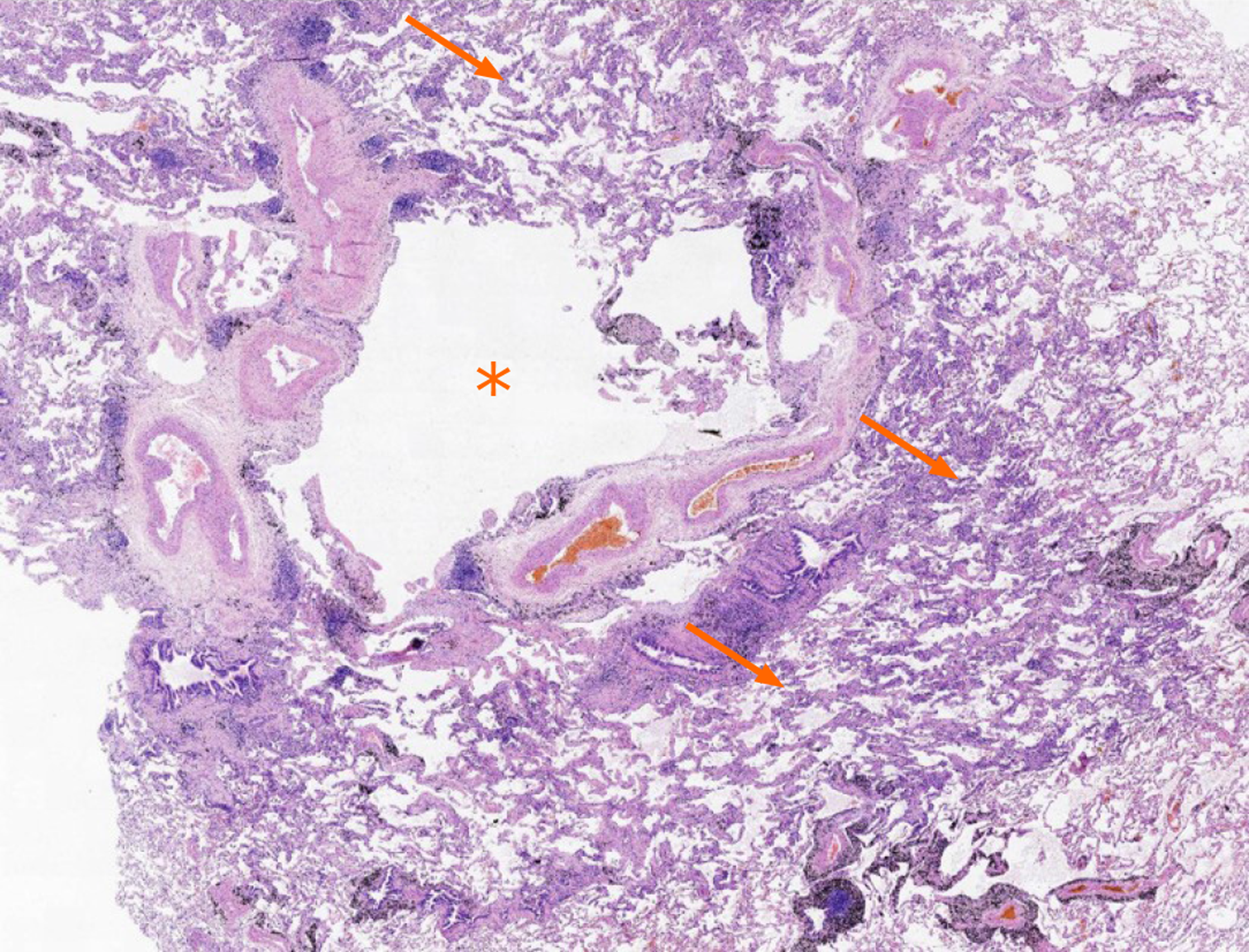
Figure 4 Histologic image (hematoxylin and eosin stain, × 2) demonstrating a part of the bulla (asterisk) and lepidic growth of adenocarcinoma (arrows).
- Citation: Meng SS, Wang SD, Zhang YY, Wang J. Lung cancer from a focal bulla into thin-walled adenocarcinoma with ground glass opacity — an observation for more than 10 years: A case report. World J Clin Cases 2020; 8(11): 2312-2317
- URL: https://www.wjgnet.com/2307-8960/full/v8/i11/2312.htm
- DOI: https://dx.doi.org/10.12998/wjcc.v8.i11.2312


