©The Author(s) 2020.
World J Clin Cases. May 26, 2020; 8(10): 2028-2037
Published online May 26, 2020. doi: 10.12998/wjcc.v8.i10.2028
Published online May 26, 2020. doi: 10.12998/wjcc.v8.i10.2028
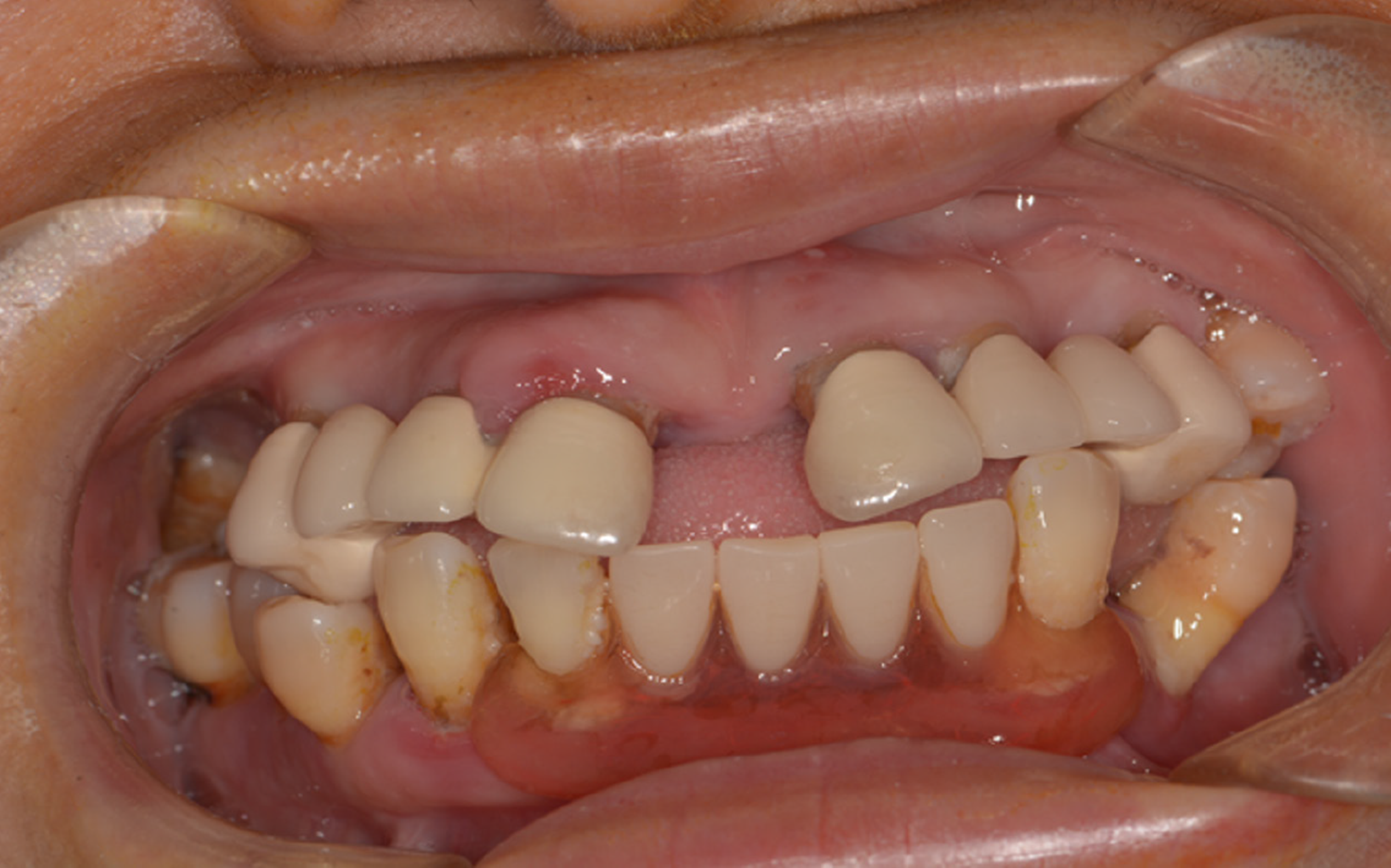
Figure 1 Intraoral photo of patient before operation.
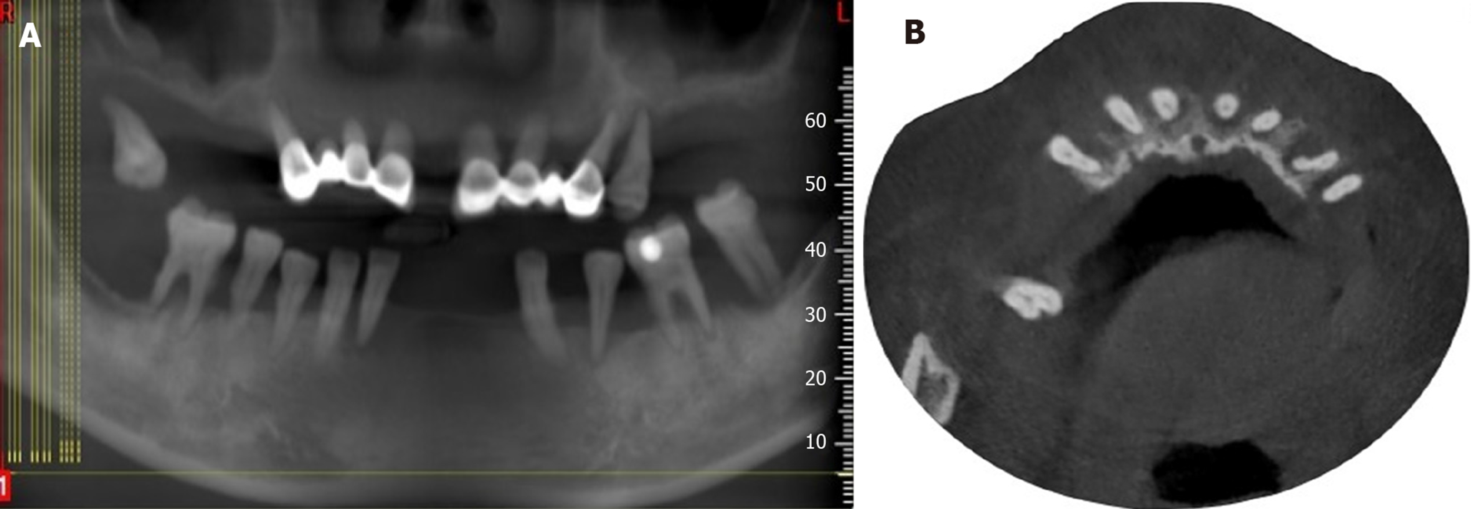
Figure 2 Image examinations before operation.
A: Stomatographic tomography of patient before operation; B: Cone beam computed tomography image of patient before operation.
Figure 3 Intraoral photo during initial surgery.
A: Buccal rectangular window at maxillary sinus lateral wall; B: Four Osstem mini implants in the maxillary bone.
Figure 4 Postoperative X-ray showing that the temporary implants were placed properly.
Figure 5 The examinations during permanent implant surgery in mandibular bond.
A: Permanent implants in the mandibular bone; B: Postoperative X-ray of implants with model transfer levers.
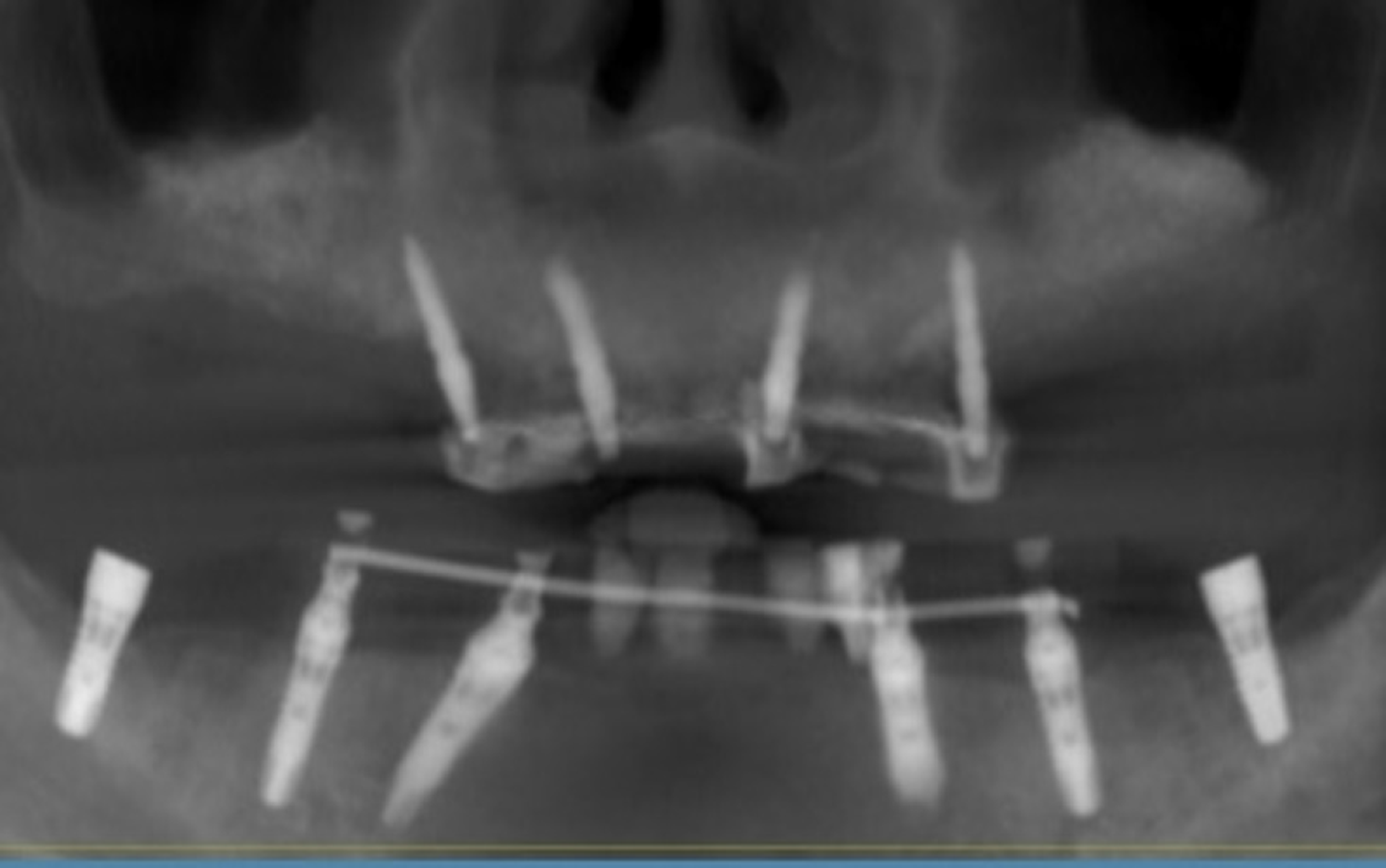
Figure 6 The examination during permanent implant surgery in maxillary bond.
A: Permanent implants in the maxillary bone during surgery; B: X-ray showing all permanent implants were placed properly.
Figure 7 Intraoral photo after full-mouth temporary restoration.
A: Temporary CAD/CAM full mouth prosthesis; B: Gingival condition after the temporary restoration was removed.
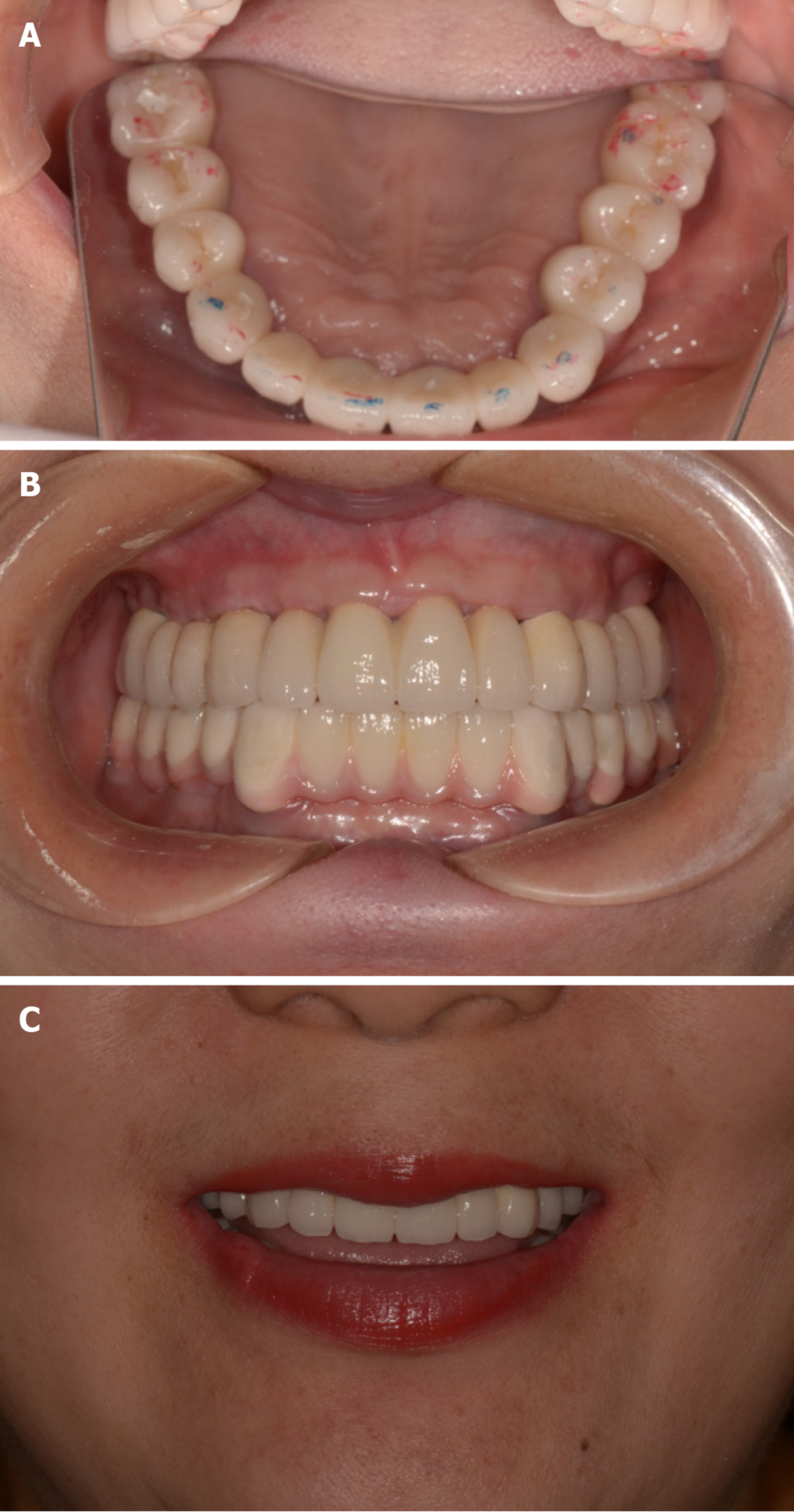
Figure 8 Intraoral photo of permanent outcomes.
A: Maxillary and mandibular restorations after the occlusal adjustment; B: Intraoral photo of zirconia prosthesis; C: Patient’s outward appearance and smile after the final procedure.
- Citation: Wang SH, Ni WC, Wang RF. Treating severe periodontitis with staged load applied implant restoration: A case report. World J Clin Cases 2020; 8(10): 2028-2037
- URL: https://www.wjgnet.com/2307-8960/full/v8/i10/2028.htm
- DOI: https://dx.doi.org/10.12998/wjcc.v8.i10.2028


