Copyright
©The Author(s) 2019.
World J Clin Cases. Mar 6, 2019; 7(5): 676-683
Published online Mar 6, 2019. doi: 10.12998/wjcc.v7.i5.676
Published online Mar 6, 2019. doi: 10.12998/wjcc.v7.i5.676
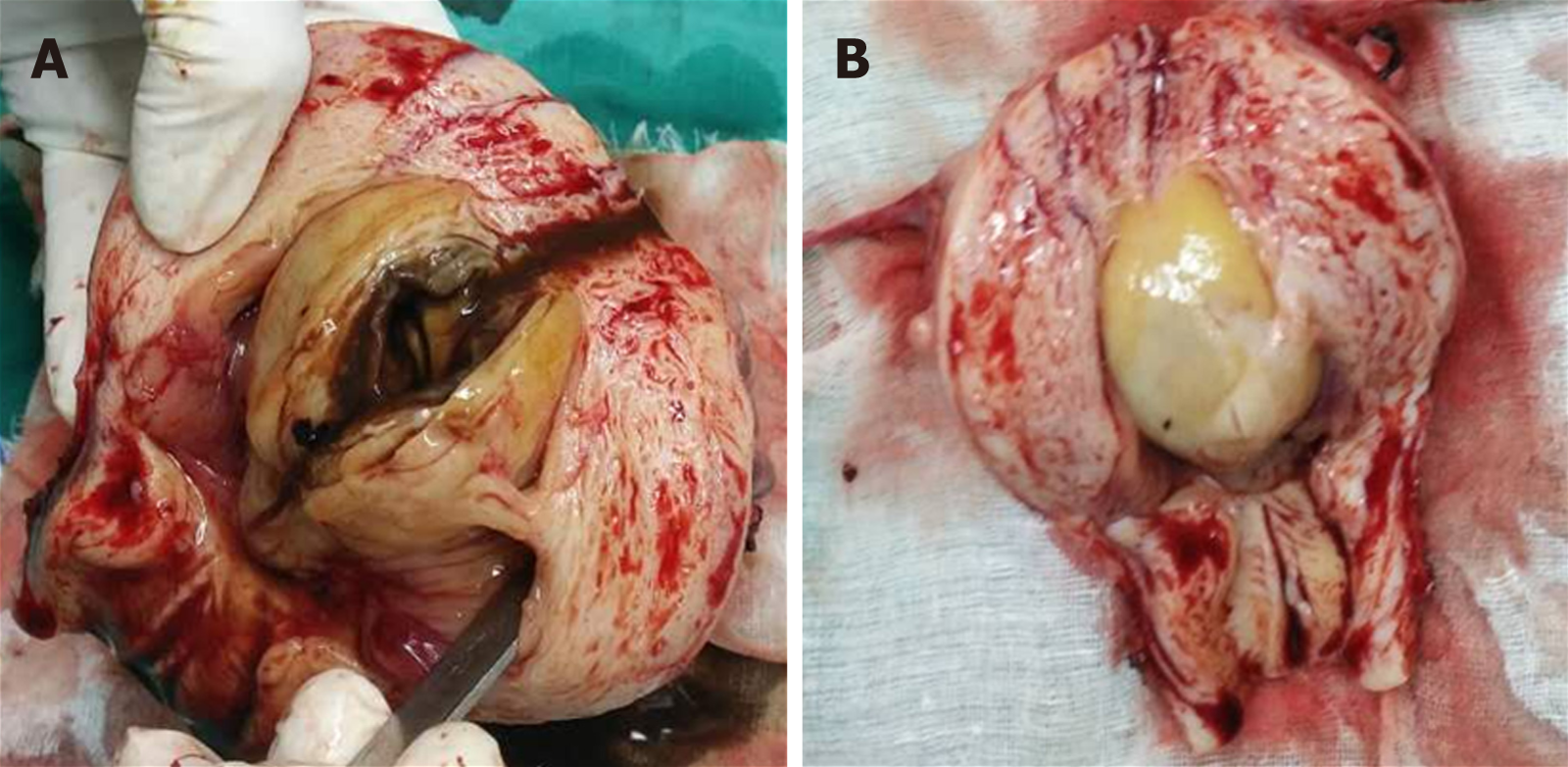
Figure 1 Gross display of intrauterine cystic adenomyosis.
A: The cyst contained chocolate-like liquid; B: After the uterus was opened, a cystic mass with a size of about 5 cm × 4 cm × 4 cm was found in the uterus, attached to the posterior wall with a pedicle with a smooth and yellow surface.
Figure 2 Ultrasonic images of intrauterine cystic adenomyosis.
Regularly shaped lesions with a high-level echo and strong echo halo around the lesions can be seen in the uterine cavity, and cysts can be seen in the polyp. Color Doppler can display the typical single blood flow signal supplying the endometrial polyps. A: Case 1; B: Case 2.
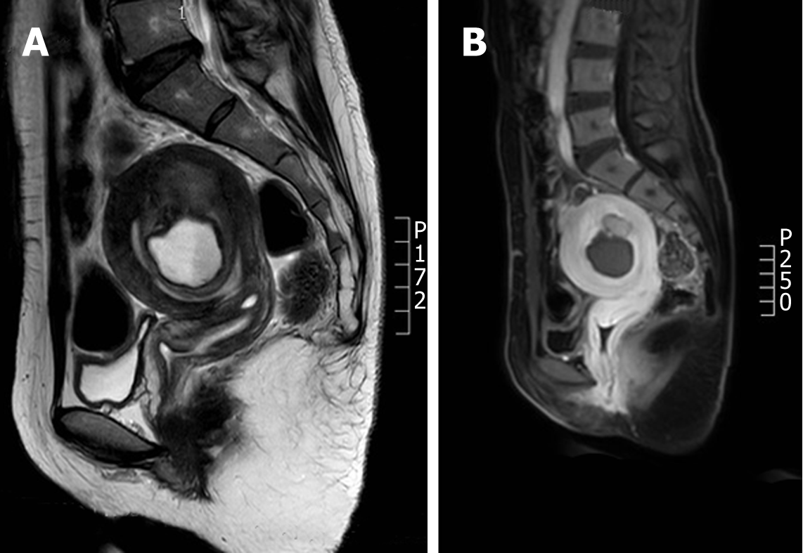
Figure 3 Magnetic resonance images of cystic adenomyosis (hyperintensity in T1 weighted image, moderate to high intensity in T2 weighted image, and low intensity in the edge).
A: Case 1; B: Case 2.
- Citation: Fan YY, Liu YN, Li J, Fu Y. Intrauterine cystic adenomyosis: Report of two cases. World J Clin Cases 2019; 7(5): 676-683
- URL: https://www.wjgnet.com/2307-8960/full/v7/i5/676.htm
- DOI: https://dx.doi.org/10.12998/wjcc.v7.i5.676


