Copyright
©The Author(s) 2019.
World J Clin Cases. Mar 6, 2019; 7(5): 642-649
Published online Mar 6, 2019. doi: 10.12998/wjcc.v7.i5.642
Published online Mar 6, 2019. doi: 10.12998/wjcc.v7.i5.642
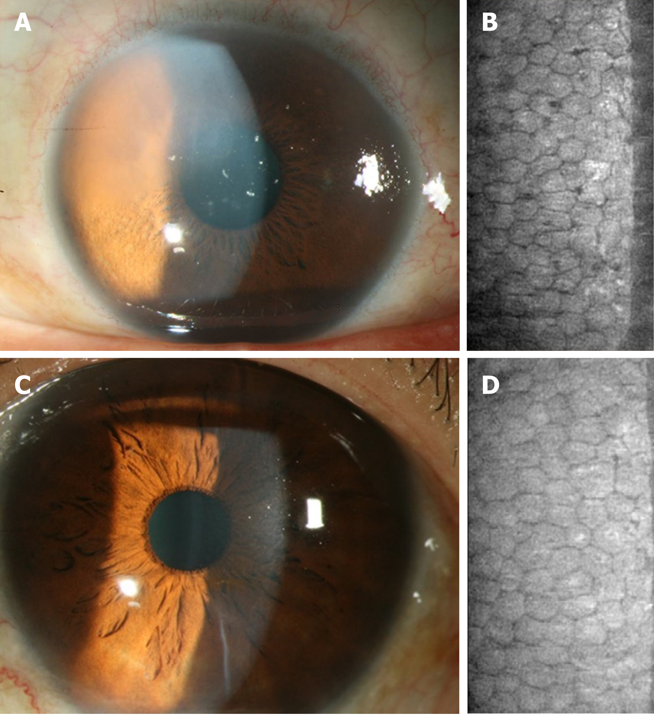
Figure 1 The corneal endothelium condition before and after cataract surgery in the left eye of Patient 2.
A: Preoperative corneal appearance via silt-lamp biomicroscope; B: Preoperative corneal endothelial cell density via specular microscope; C: Postoperative corneal appearance via silt-lamp biomicroscope; D: Postoperative corneal endothelial cell density via specular microscope.
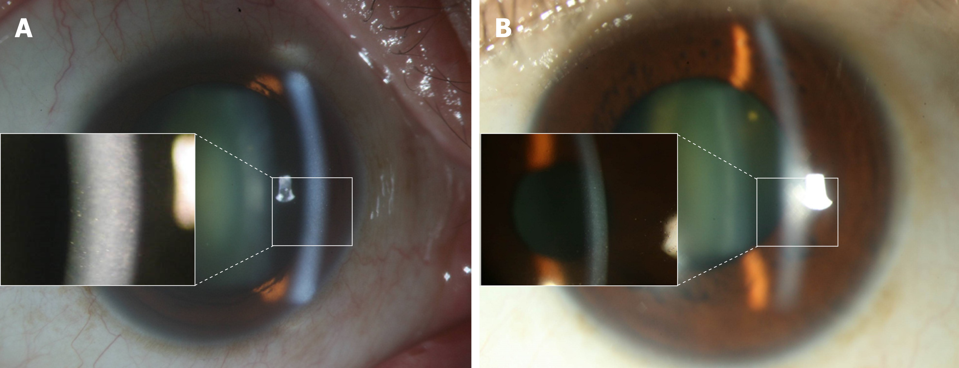
Figure 2 The appearance of Fuchs endothelial corneal dystrophy in Patient 1.
A: External eye appearance and guttate formation (left bracket) in the right eye; B: External eye appearance and guttate formation (left bracket) in the left eye.
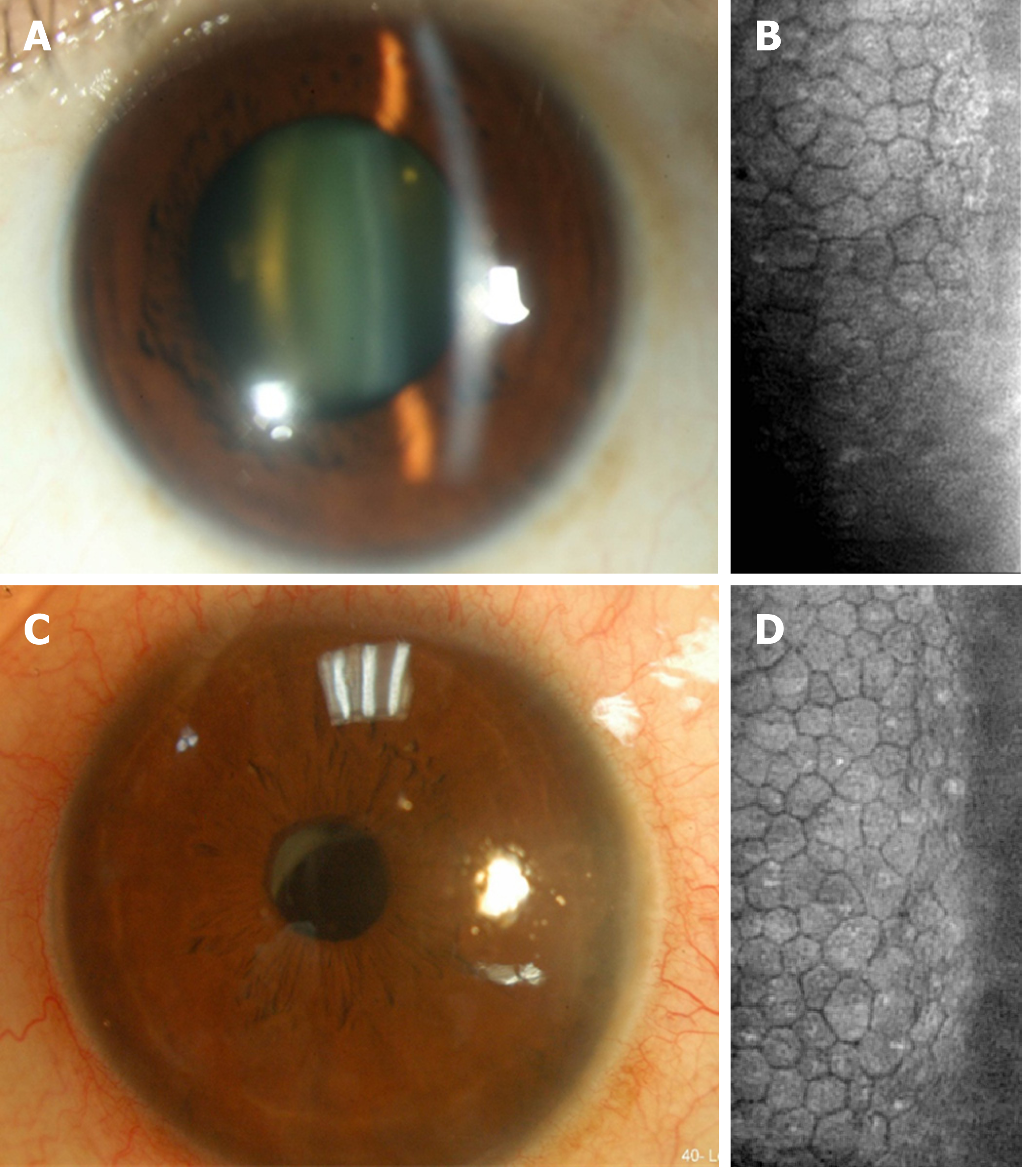
Figure 3 The corneal endothelium condition before and after cataract surgery in the left eye of Patient 1.
A: Preoperative corneal appearance via silt-lamp biomicroscope; B: Preoperative corneal endothelial cell density via specular microscope; C: Postoperative corneal appearance via silt-lamp biomicroscope; D: Postoperative corneal endothelial cell density via specular microscope.
- Citation: Lee CY, Chen HT, Hsueh YJ, Chen HC, Huang CC, Meir YJJ, Cheng CM, Wu WC. Perioperative topical ascorbic acid for the prevention of phacoemulsification-related corneal endothelial damage: Two case reports and review of literature. World J Clin Cases 2019; 7(5): 642-649
- URL: https://www.wjgnet.com/2307-8960/full/v7/i5/642.htm
- DOI: https://dx.doi.org/10.12998/wjcc.v7.i5.642


