©The Author(s) 2019.
World J Clin Cases. Feb 6, 2019; 7(3): 373-381
Published online Feb 6, 2019. doi: 10.12998/wjcc.v7.i3.373
Published online Feb 6, 2019. doi: 10.12998/wjcc.v7.i3.373
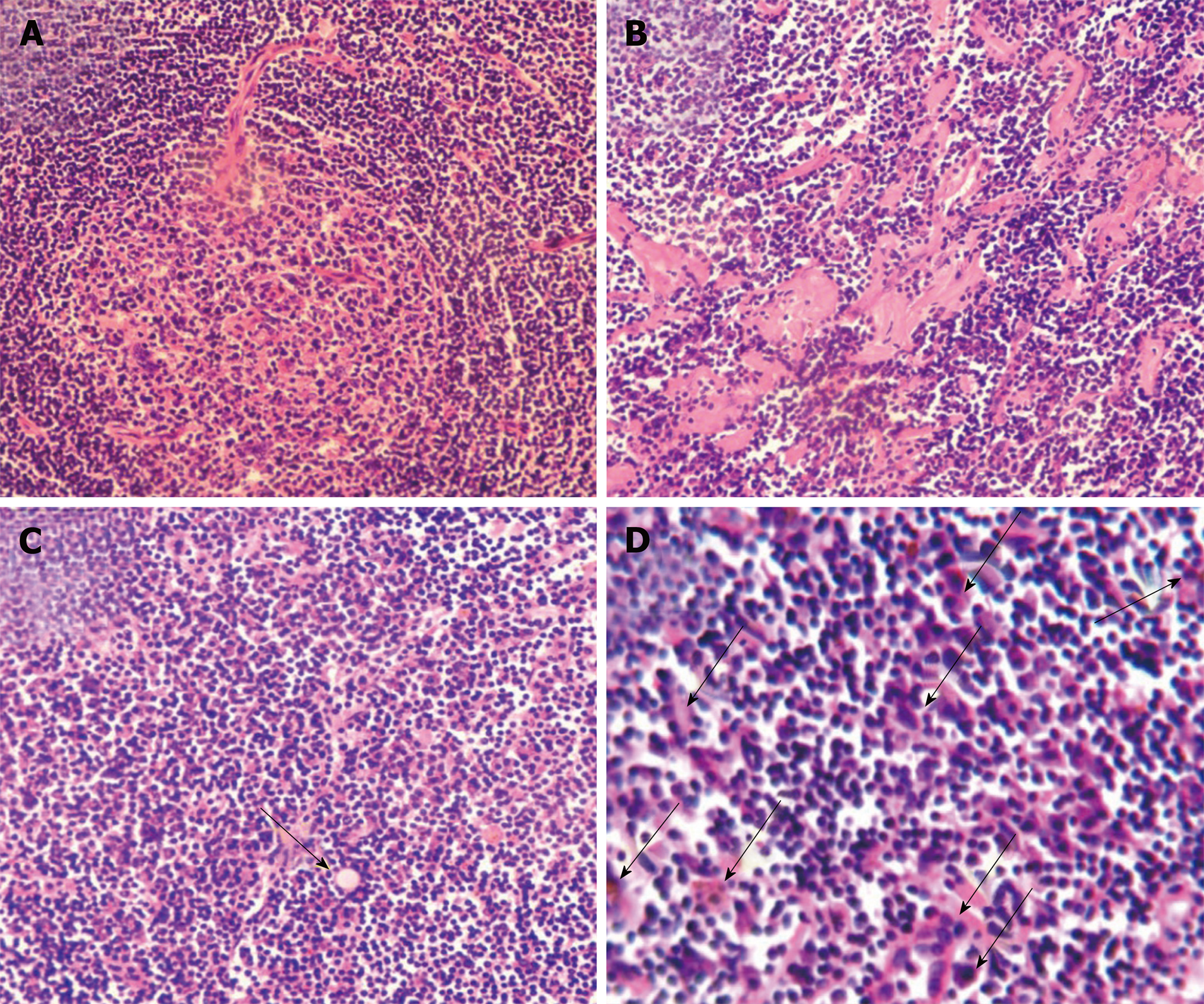
Figure 1 Major pathohistological features of mixed type Castleman disease.
Tissue sections (5 μm) were stained with hematoxylin and eosin. Histological characteristics in representative images include hyperplasia of follicular lymphoids concentrically layered around vascularized and degenerative germinal centers and shortage of lymphatic sinuses (A), hyaline degeneration (B), existence of Russell’s body (arrow) (C), and abundant proliferating plasma cells (arrows) (C and D). Magnification, × 200 (A-C) and × 400 (D).
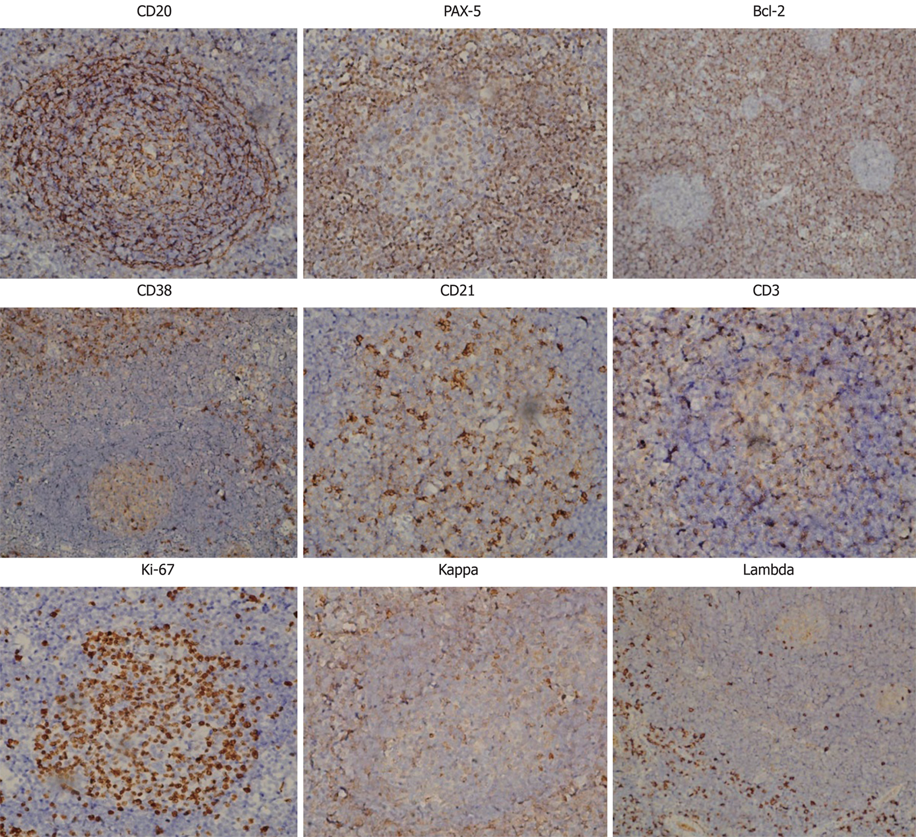
Figure 2 Immunohistochemistry.
A panel of antibodies as indicated were used to immunostain the sections, which were subsequently counterstained with hematoxylin. Magnification, × 200. PAX-5: Paired box protein-5; Bcl-2: B-cell lymphoma 2.
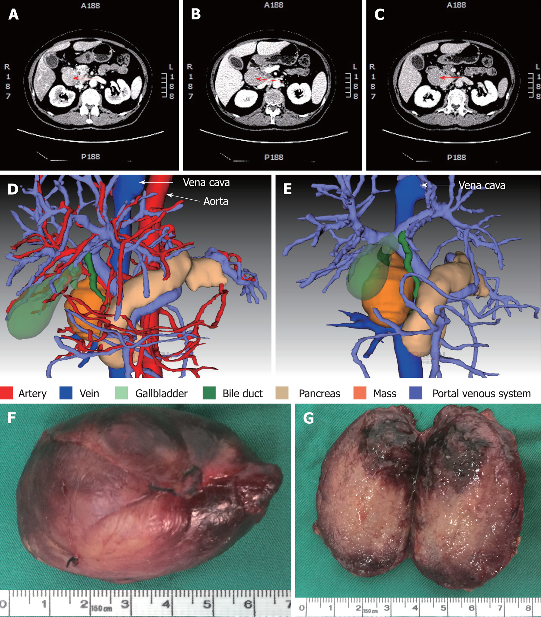
Figure 3 Computed tomography scan, three-dimensional visualization model and the mass.
A-C: Representative images are from enhanced multi-phase abdominal computed tomography scans at the arterial (A), portal venous (B) and equilibrium (C) phases. Arrows point to the mass. D and E: Three-dimensional images were generated to show the adjacent relationship between the mass and adjacent organs and tissues. F: The excised mass. G: The cutting surface.
- Citation: Zhai B, Ren HY, Li WD, Reddy S, Zhang SJ, Sun XY. Castleman disease presenting with jaundice: A case report and review of literature. World J Clin Cases 2019; 7(3): 373-381
- URL: https://www.wjgnet.com/2307-8960/full/v7/i3/373.htm
- DOI: https://dx.doi.org/10.12998/wjcc.v7.i3.373


