©The Author(s) 2019.
World J Clin Cases. Dec 26, 2019; 7(24): 4391-4397
Published online Dec 26, 2019. doi: 10.12998/wjcc.v7.i24.4391
Published online Dec 26, 2019. doi: 10.12998/wjcc.v7.i24.4391
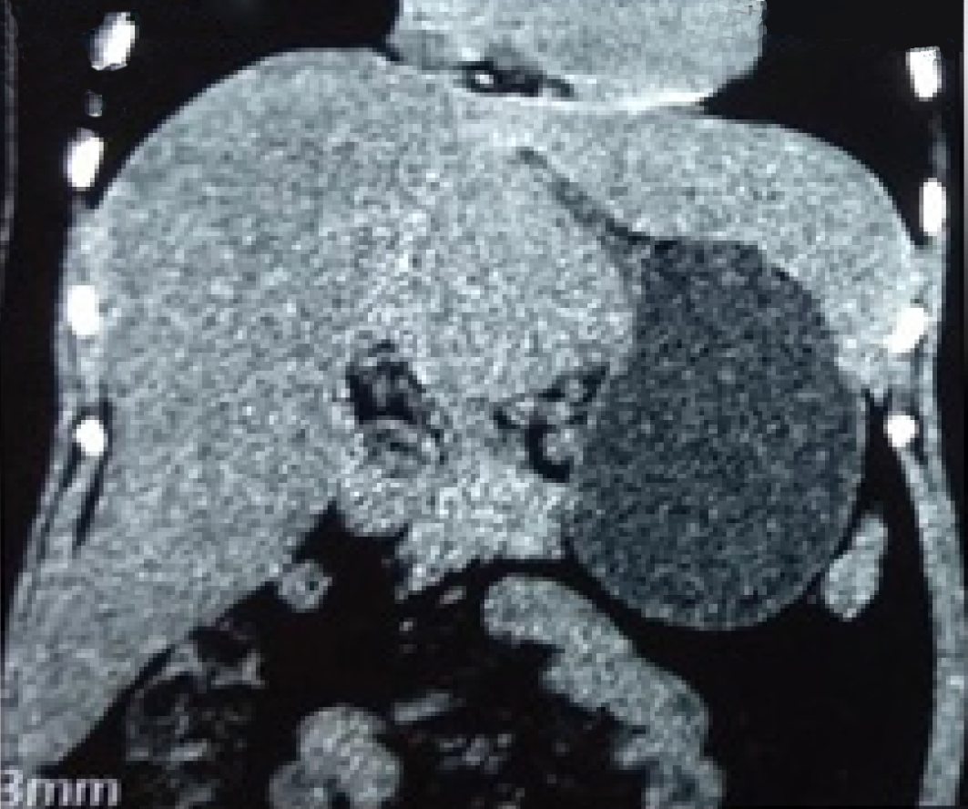
Figure 1 Computed tomography image showing circular soft tissue nodule in the hepatic-gastric space.
Figure 2 Enhanced computed tomography images of the patient.
A: Plain scan; B: Arterial phase; C: Venous phase.
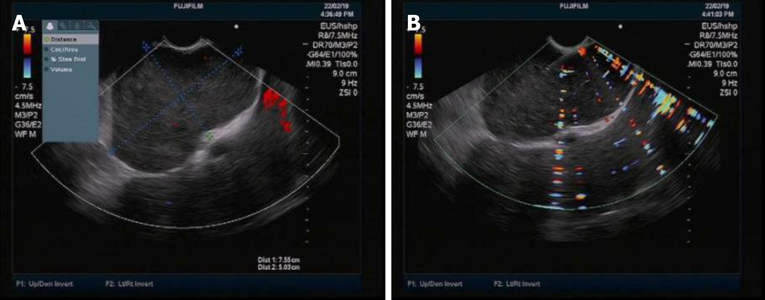
Figure 3 Endoscopic ultrasound images of the patient.
A: Endoscopic ultrasound revealed low echoes in the hepatic-gastric space with scattered star-shaped high echoes. Color Doppler revealed absence of blood flow signal; B: Endoscopic ultrasound guided fine needle aspiration.
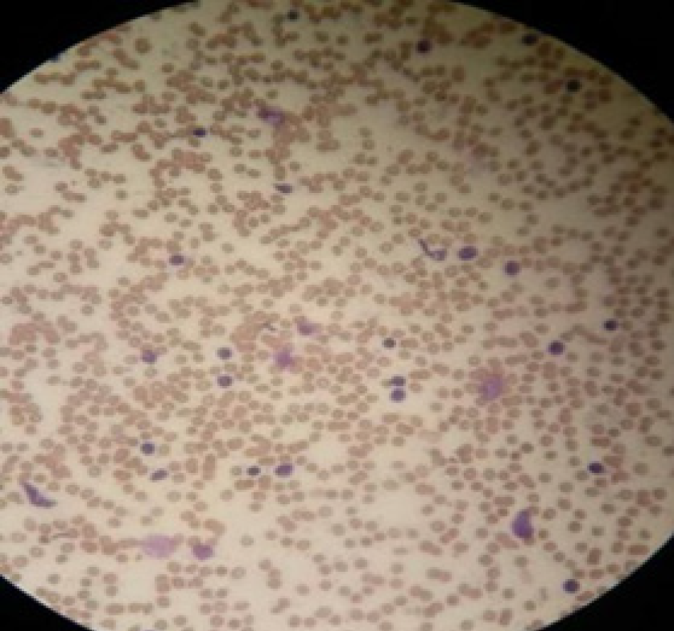
Figure 4 Diff-Quik staining showed a large number of scattered lymphocytes with normal morphology under the background of red blood cells.
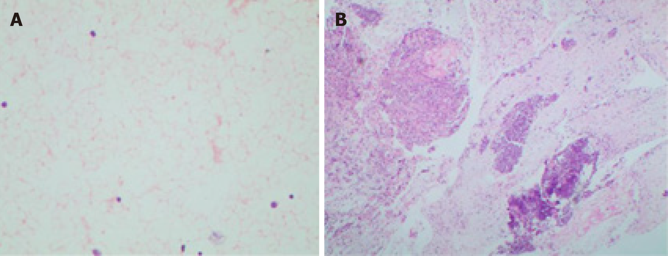
Figure 5 Hematoxylin eosin staining of the smear and tissue.
A: Hematoxylin eosin staining of the smear: Scattered lymphocytes with normal morphology; B: Hematoxylin eosin staining of the tissue: A large number of lymphocytes with normal morphology, dominated by B cells.
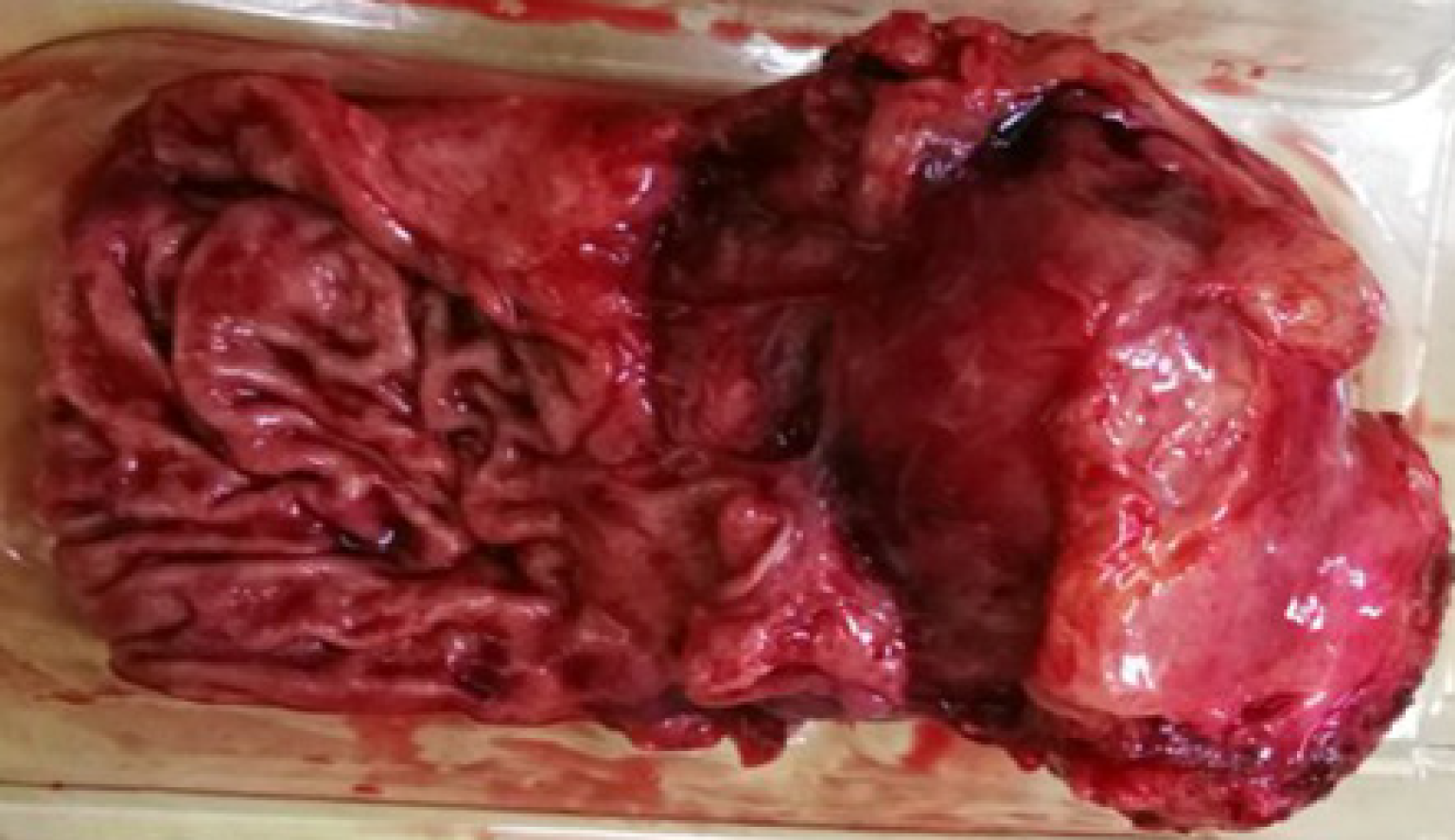
Figure 6 Surgical specimens.
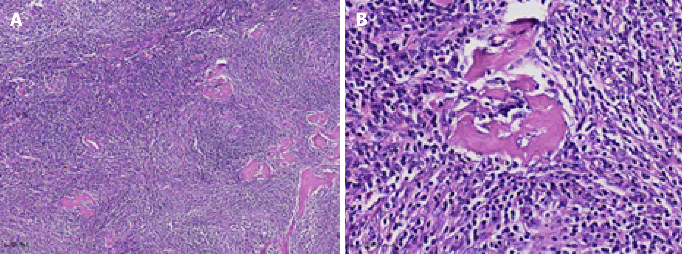
Figure 7 Postoperative pathological hematoxylin eosin staining.
A: × 100; B: × 400.
- Citation: Xu XY, Liu XQ, Du HW, Liu JH. Castleman disease in the hepatic-gastric space: A case report. World J Clin Cases 2019; 7(24): 4391-4397
- URL: https://www.wjgnet.com/2307-8960/full/v7/i24/4391.htm
- DOI: https://dx.doi.org/10.12998/wjcc.v7.i24.4391


