©The Author(s) 2019.
World J Clin Cases. Dec 26, 2019; 7(24): 4384-4390
Published online Dec 26, 2019. doi: 10.12998/wjcc.v7.i24.4384
Published online Dec 26, 2019. doi: 10.12998/wjcc.v7.i24.4384
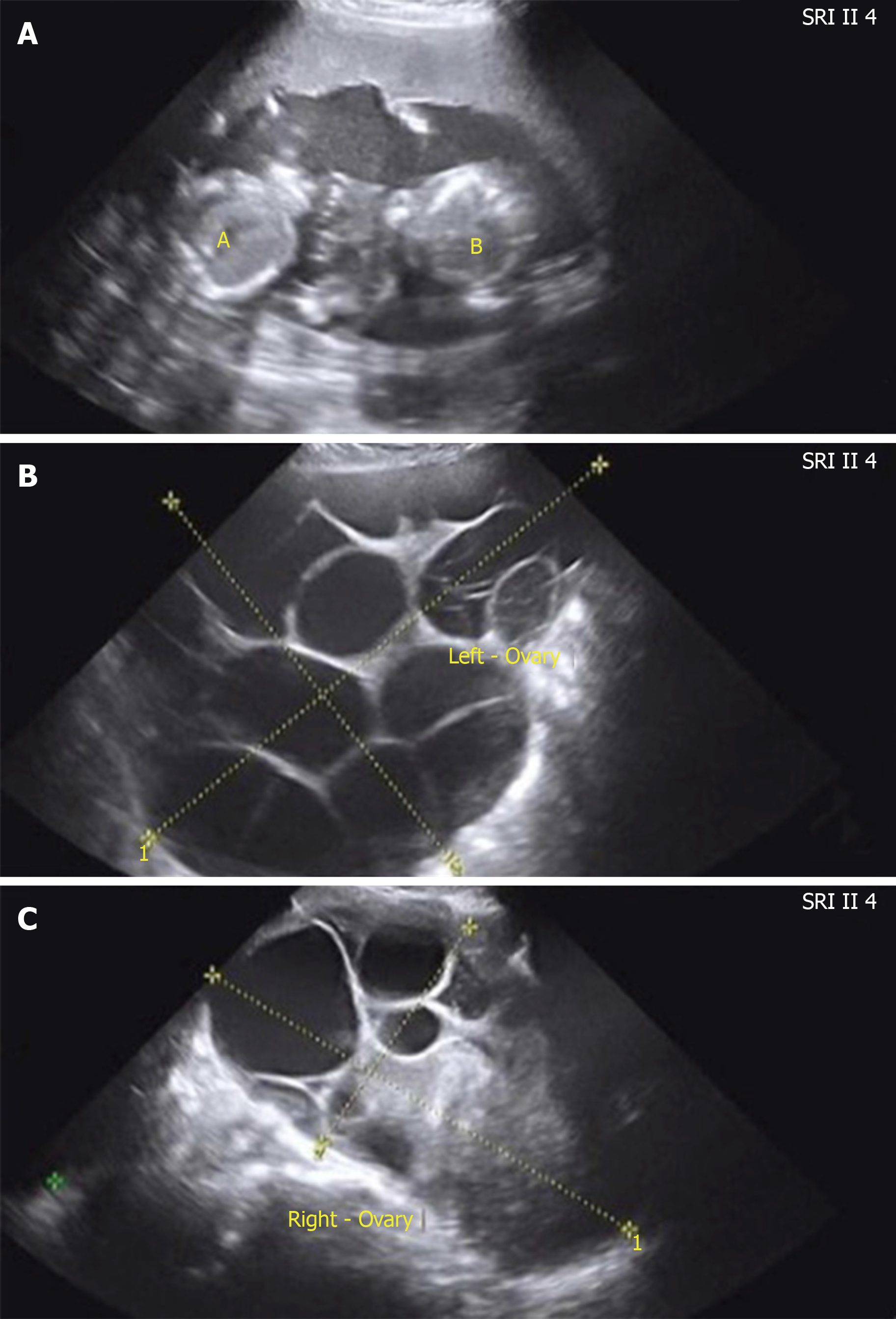
Figure 1 Abdominal ultrasonography of the fetus, ovary, and ascites in case 2.
A: Ultrasonography of the fetus showed a twin pregnancy at the 17th gestational week; B: Ultrasonography of the left ovary showed that the size of the left ovary was 28.46 cm × 12.36 cm; C: Ultrasonography of the right ovary showed that the size of the right ovary was right 15.01 cm × 11.27 cm.
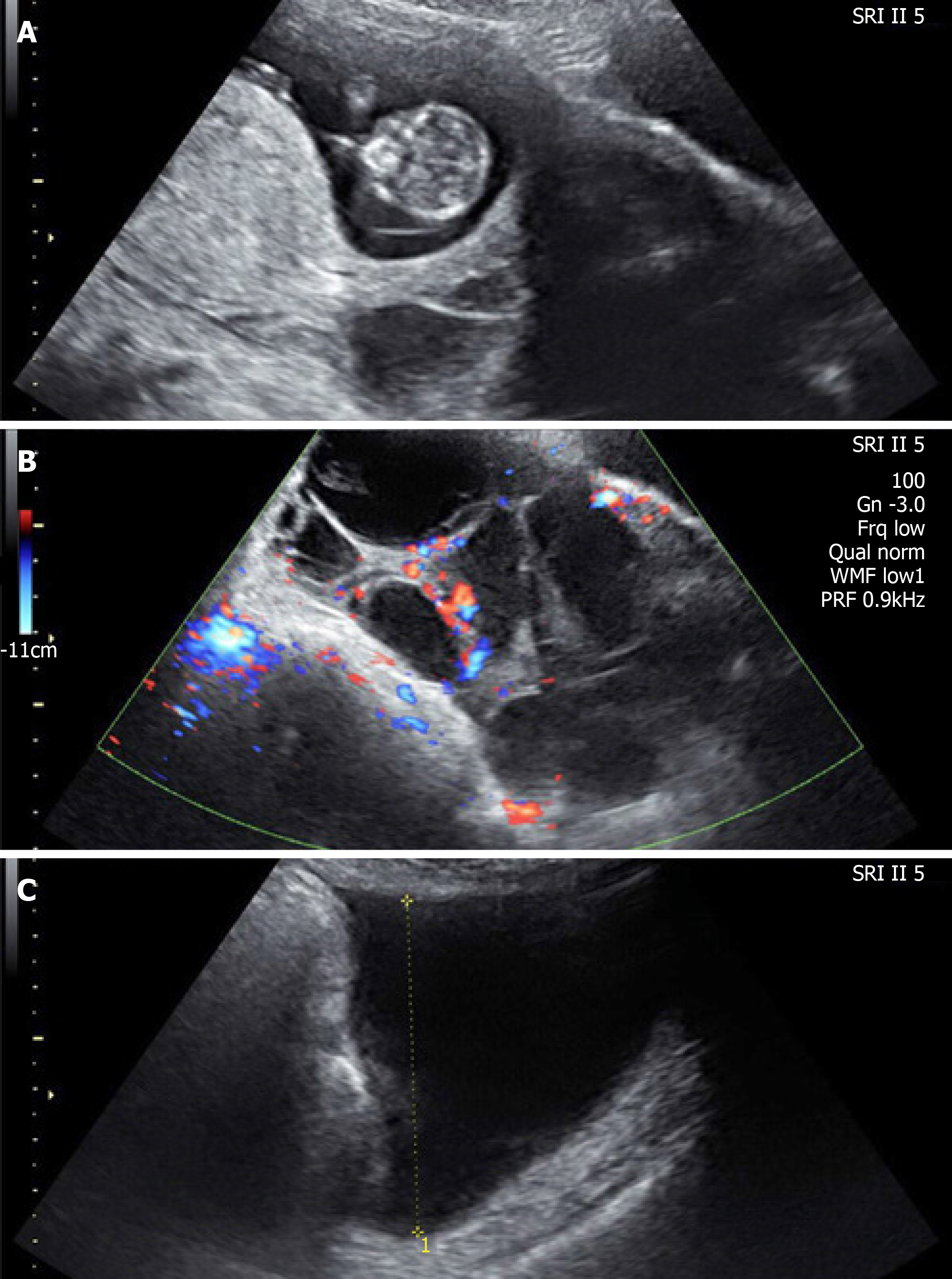
Figure 2 Abdominal ultrasonography of the fetus, ovary, and ascites in case 1.
A: Ultrasonography of the fetus showed a single live fetus at 12 + 5 gestational weeks; B: Ultrasonography of the ovaries showed bilateral ovarian enlargement (the size of the left ovary was 14.92 cm × 7.98 cm and the size of the right ovary was 13.33 cm × 8.11 cm) and bilateral cystic adnexal masses (left 4.91 cm × 4.33 cm, right 3.86 cm × 2.97 cm); C: Ultrasonography showed massive ascites.
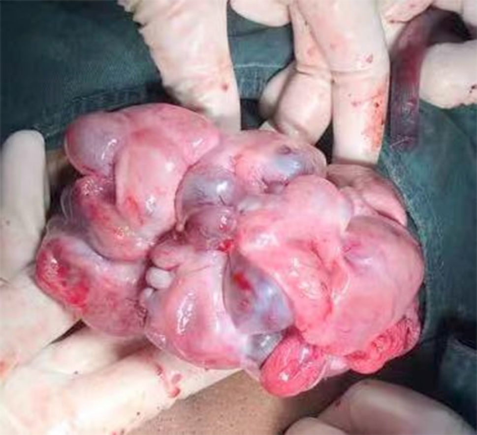
Figure 3 Enlarged ovary during cesarean section in case 2.
Enlarged ovary during operation.
- Citation: Gui J, Zhang J, Xu WM, Ming L. Spontaneous ovarian hyperstimulation syndrome: Report of two cases. World J Clin Cases 2019; 7(24): 4384-4390
- URL: https://www.wjgnet.com/2307-8960/full/v7/i24/4384.htm
- DOI: https://dx.doi.org/10.12998/wjcc.v7.i24.4384


