Copyright
©The Author(s) 2019.
World J Clin Cases. Nov 6, 2019; 7(21): 3517-3523
Published online Nov 6, 2019. doi: 10.12998/wjcc.v7.i21.3517
Published online Nov 6, 2019. doi: 10.12998/wjcc.v7.i21.3517
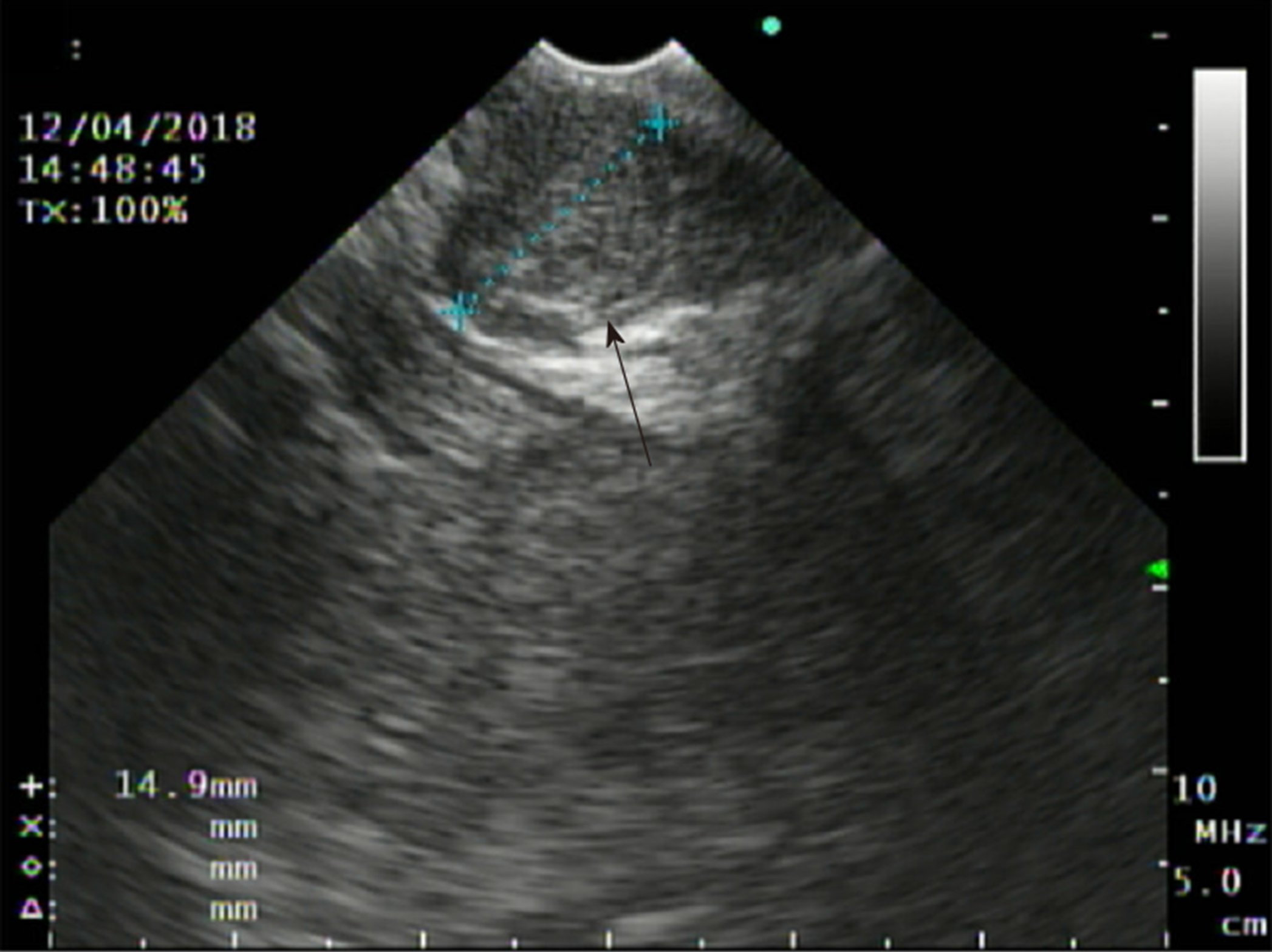
Figure 1 Endoscopic ultrasound demonstrating a well-circumscribed, hypoechoic, subepithelial antral mass.
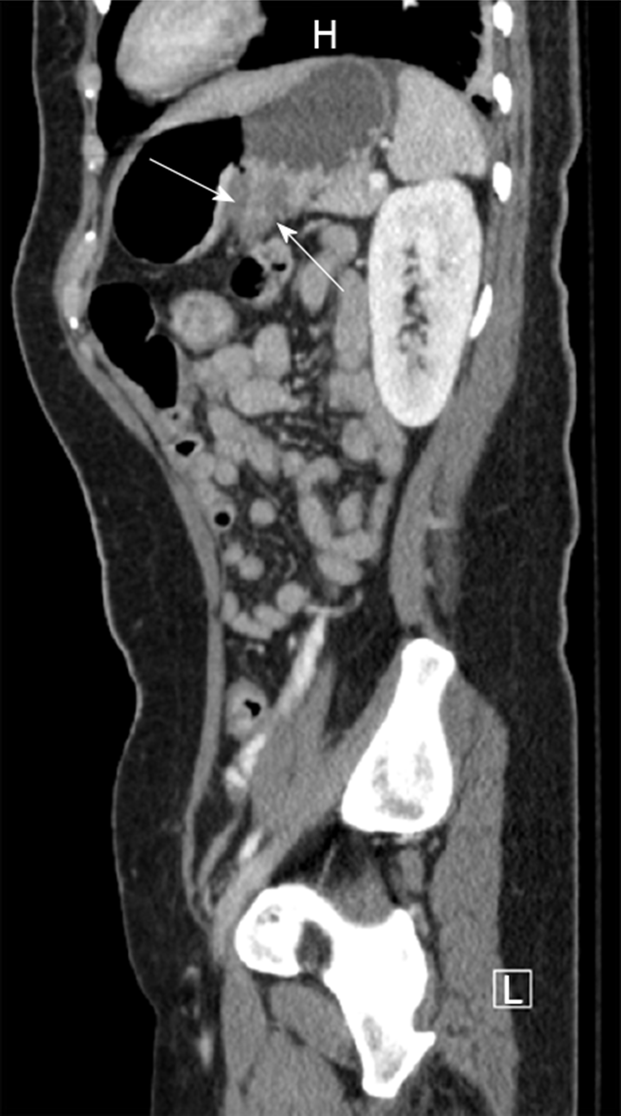
Figure 2 CT of the abdomen and pelvis, sagittal view, revealing gastro-gastric intussusception.
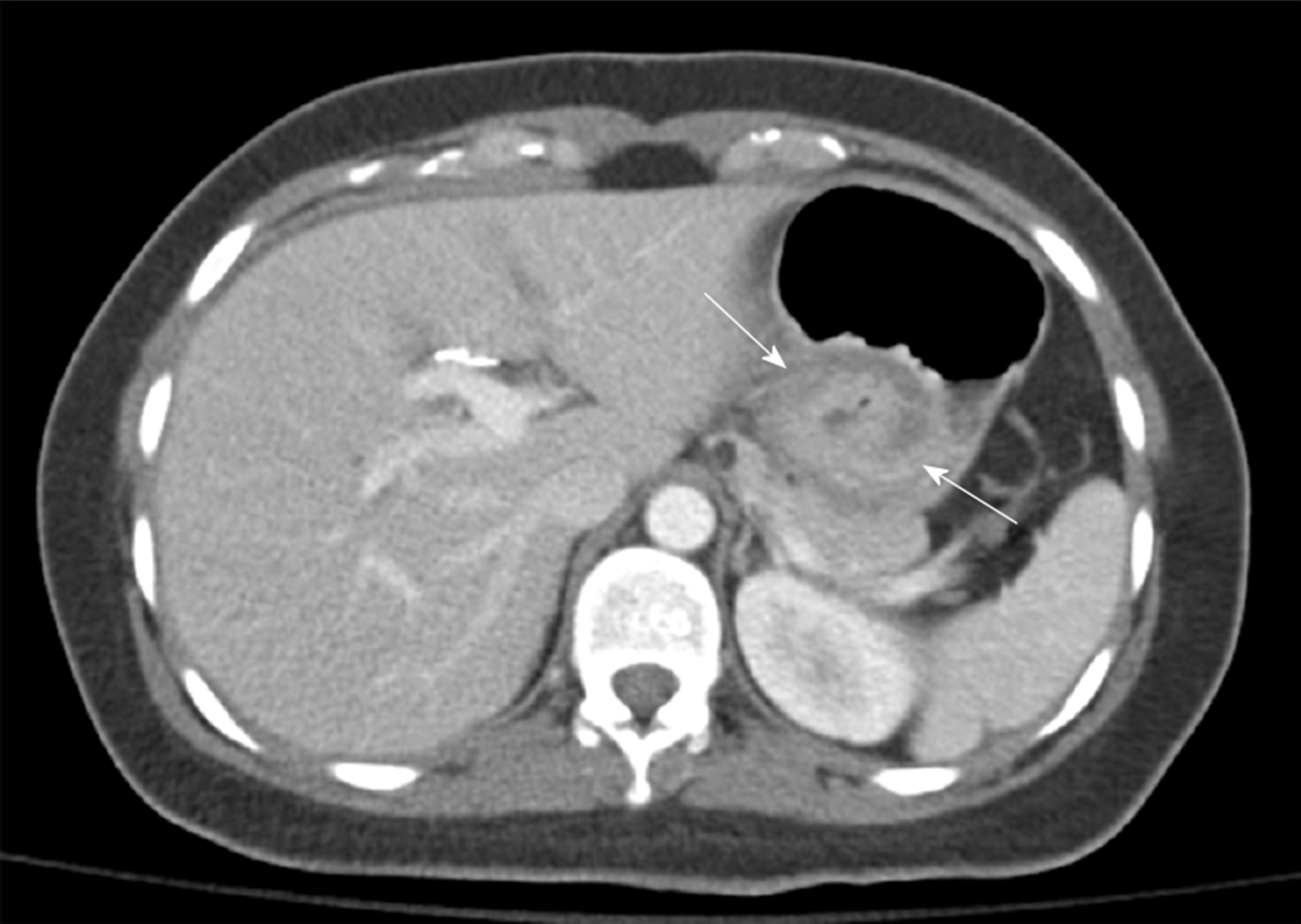
Figure 3 CT of the abdomen and pelvis, axial view, demonstrating gastro-gastric intussusception.
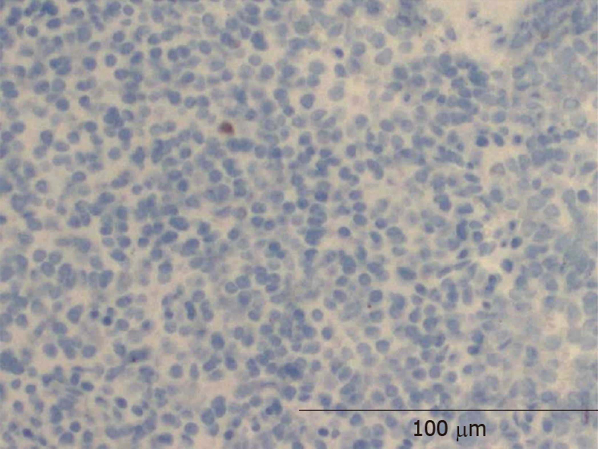
Figure 4 Histopathology showing low Ki67 staining (100x magnification).
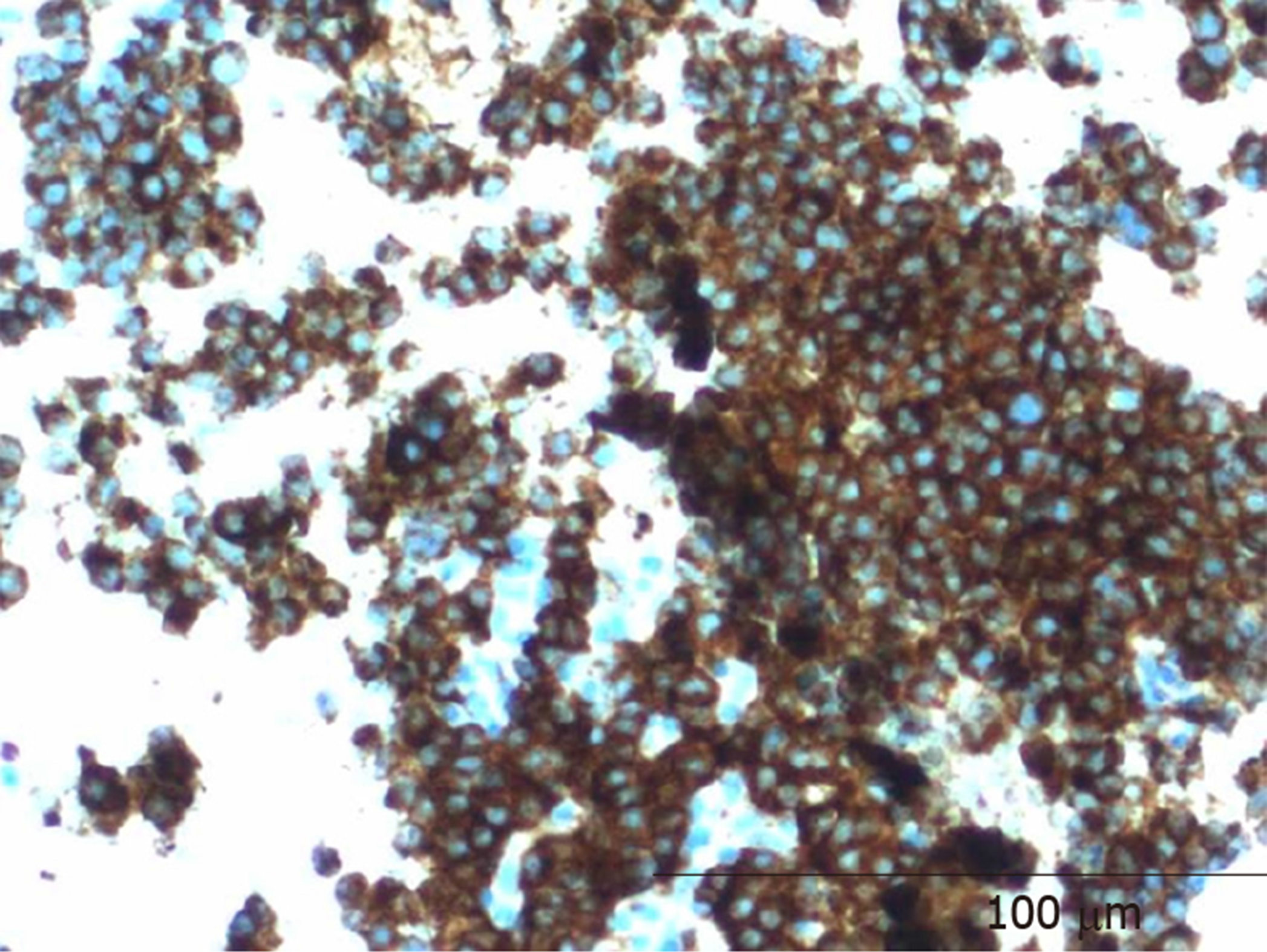
Figure 5 Histopathology showing positive synaptophysin staining (100x magnification).
- Citation: Zhornitskiy A, Le L, Tareen S, Abdullahi G, Karunasiri D, Tabibian JH. Gastro-gastric intussusception in the setting of a neuroendocrine tumor: A case report. World J Clin Cases 2019; 7(21): 3517-3523
- URL: https://www.wjgnet.com/2307-8960/full/v7/i21/3517.htm
- DOI: https://dx.doi.org/10.12998/wjcc.v7.i21.3517


