Copyright
©The Author(s) 2019.
World J Clin Cases. Oct 26, 2019; 7(20): 3322-3328
Published online Oct 26, 2019. doi: 10.12998/wjcc.v7.i20.3322
Published online Oct 26, 2019. doi: 10.12998/wjcc.v7.i20.3322
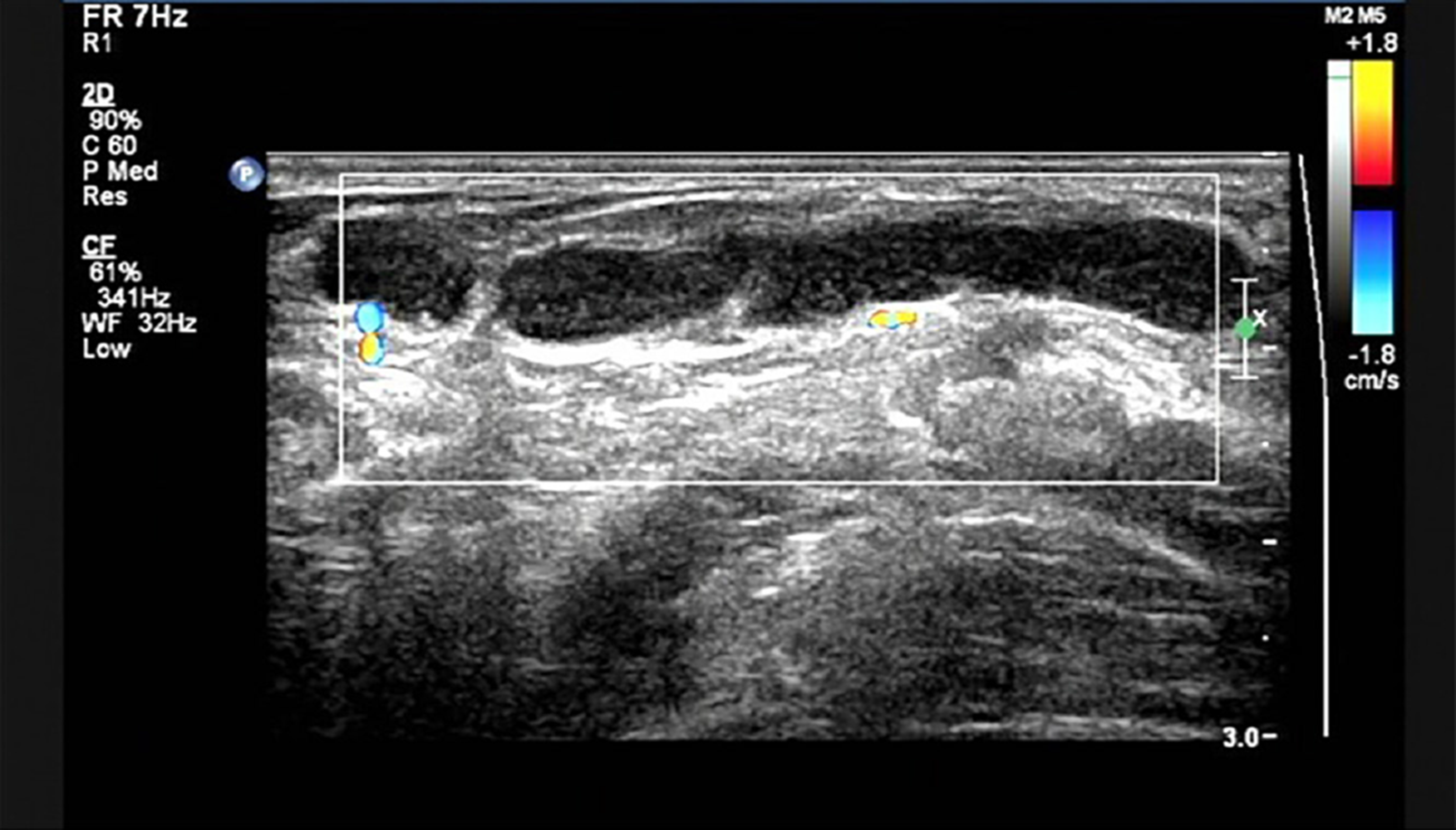
Figure 1 Ultrasound scan showing one of the subcutaneous fluid-filled regions from the left vulva to pubic symphysis that was multilocular and mobile.
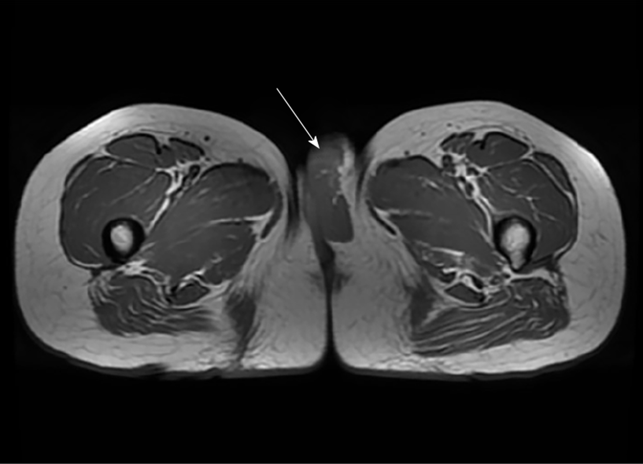
Figure 2 Pelvic enhanced nuclear magnetic resonance scan revealing a cystic area in the left vulva, as shown by the arrow.
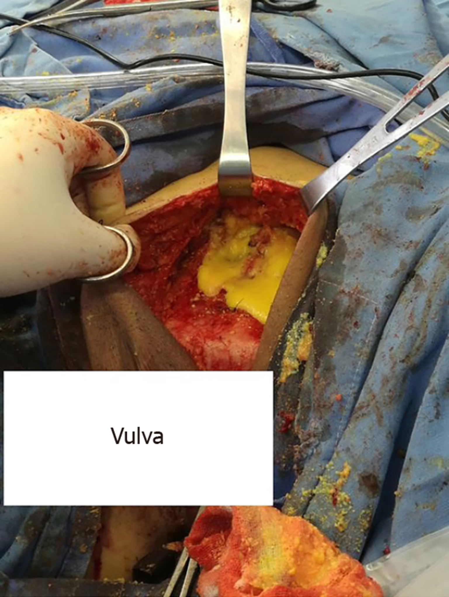
Figure 3 Yellow material that resembled bean dregs without an obvious capsule was found during surgery.
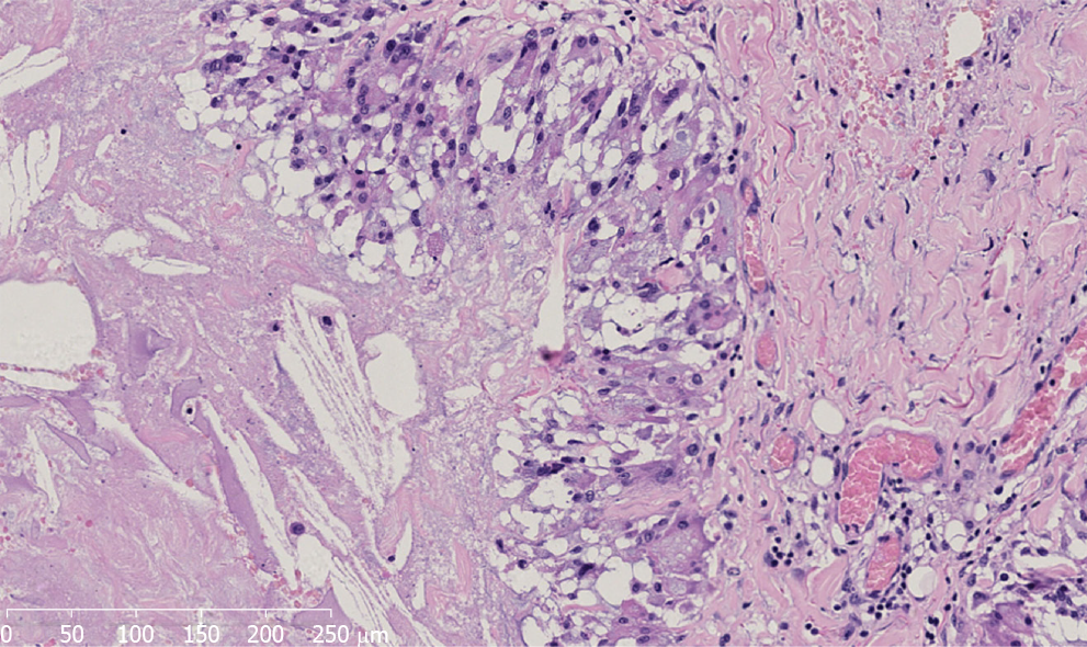
Figure 4 Microscopic appearance of the gray-white and gray-yellow tissue revealed dilated or fissured interconnected cysts with pseudopapillary structures.
The cyst wall was composed of fibrous tissue with hyaline degeneration. There are many tissue cells and foreign body giant cells in the inner wall (hematoxylin and eosin staining; original magnification: 200×).
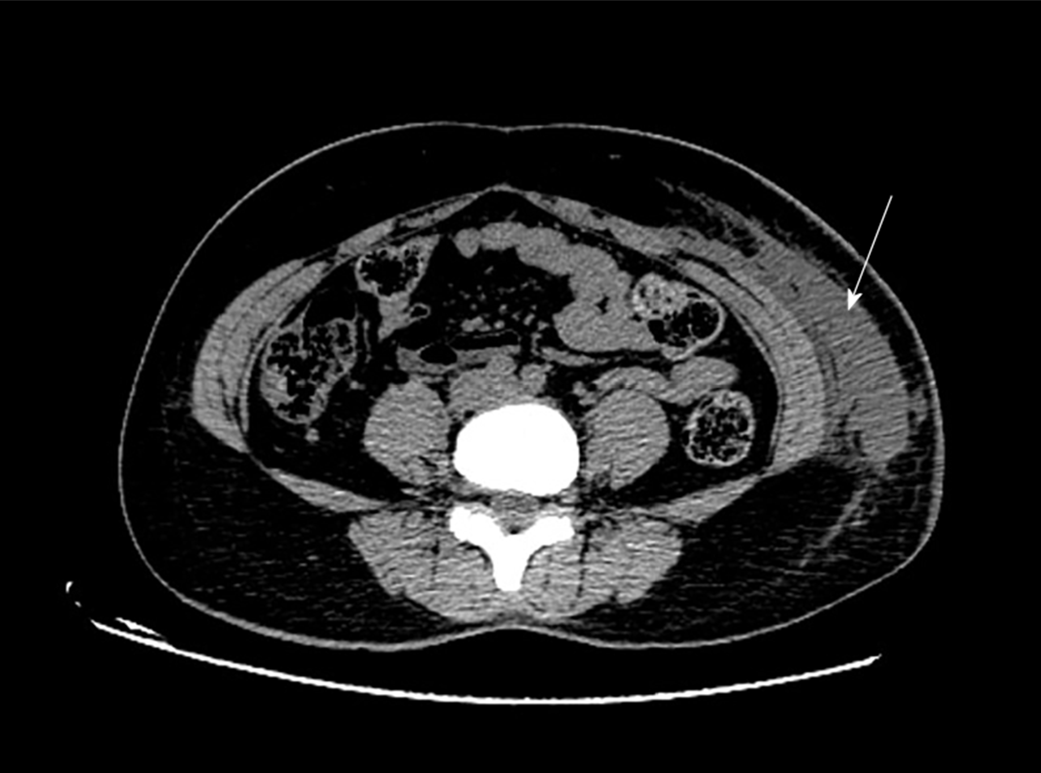
Figure 5 An abdominal computed tomography scan showed extensive infiltration and effusion from the left hypochondriac region to the vulva, as indicated by the arrow.
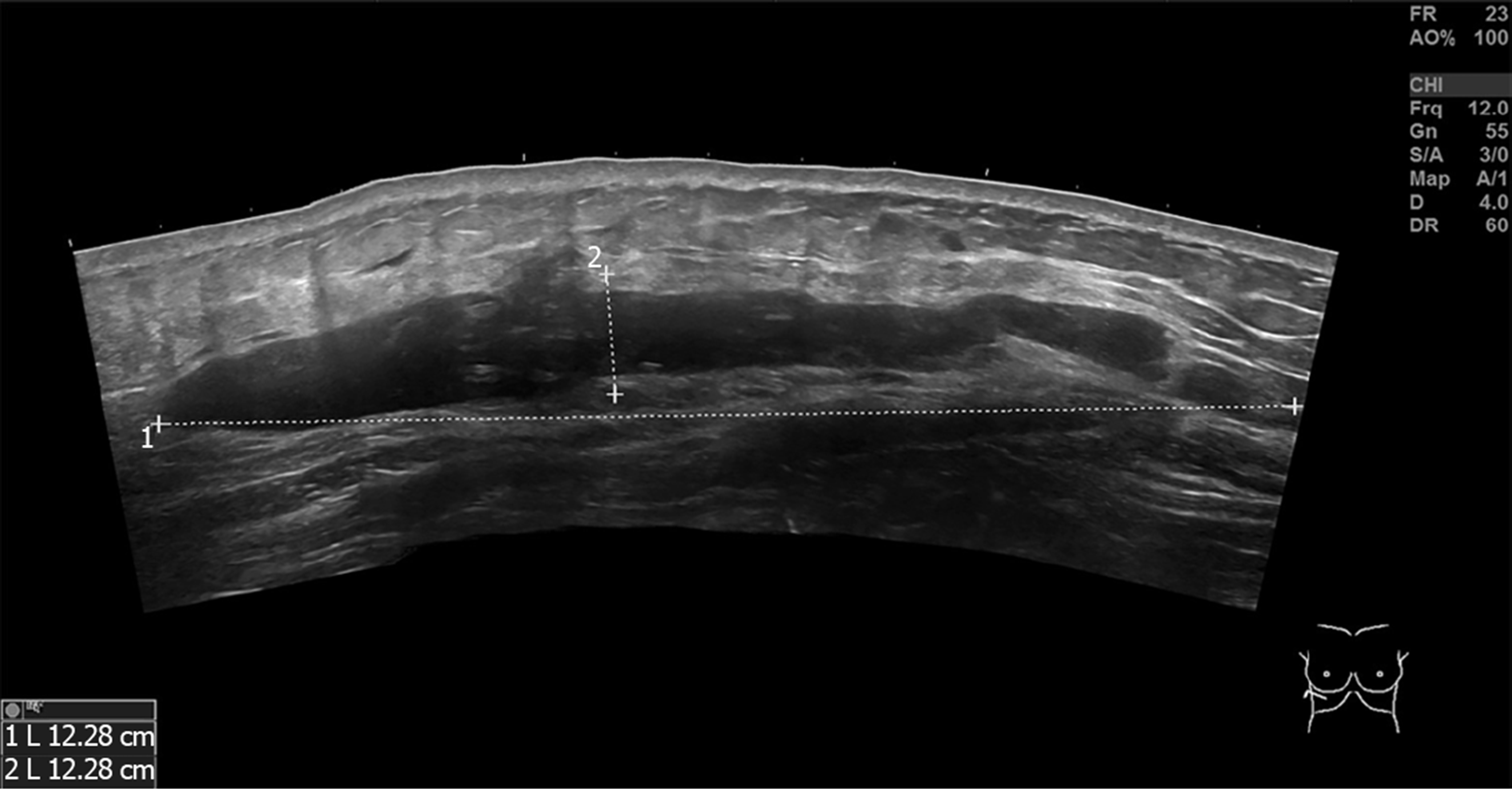
Figure 6 Ultrasonography of the bilateral hypochondriac region demonstrating that there was a hypoechoic area shaped like a bar on each side.
- Citation: Zhang MX, Li SY, Xu LL, Zhao BW, Cai XY, Wang GL. Repeated lumps and infections: A case report on breast augmentation complications. World J Clin Cases 2019; 7(20): 3322-3328
- URL: https://www.wjgnet.com/2307-8960/full/v7/i20/3322.htm
- DOI: https://dx.doi.org/10.12998/wjcc.v7.i20.3322


