Copyright
©The Author(s) 2019.
World J Clin Cases. Oct 26, 2019; 7(20): 3259-3265
Published online Oct 26, 2019. doi: 10.12998/wjcc.v7.i20.3259
Published online Oct 26, 2019. doi: 10.12998/wjcc.v7.i20.3259
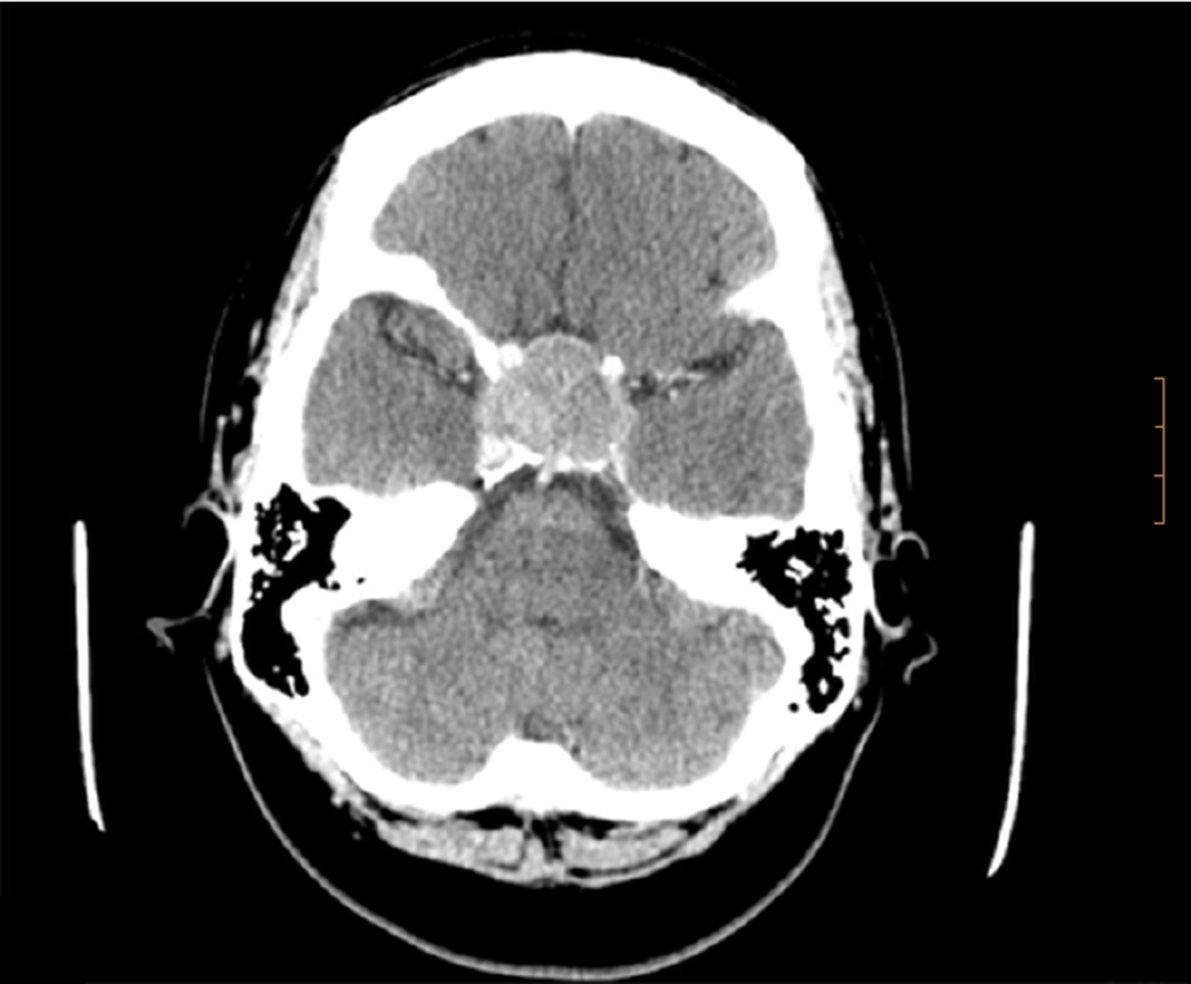
Figure 1 Axial brain computed tomography scan with contrast media administration.
Image shows enlarged pituitary gland (3.5 cm) impinging upon the optic chiasm with bone involvement of the sella. The tumour invaded the cavernous sinuses. Homogeneous enhancement with high density focus that suggests hemorrhagic infarction of the tumor.
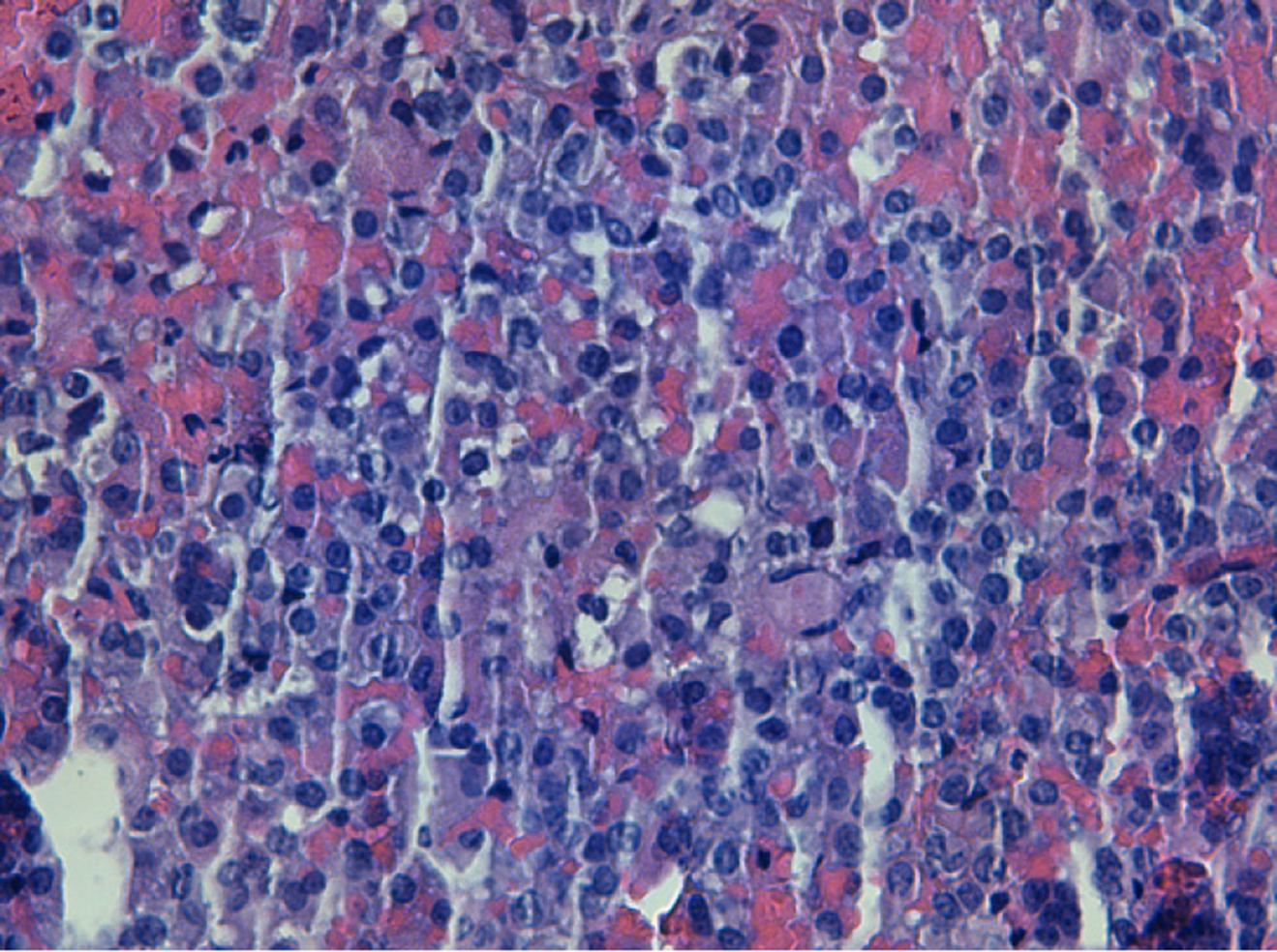
Figure 2 Microscopic findings of the pituitary adenoma.
It shows proliferation of ovoid cells, monotonous, with irregular chromatin, without showing cytological atypia or mitosis figures (× 200).
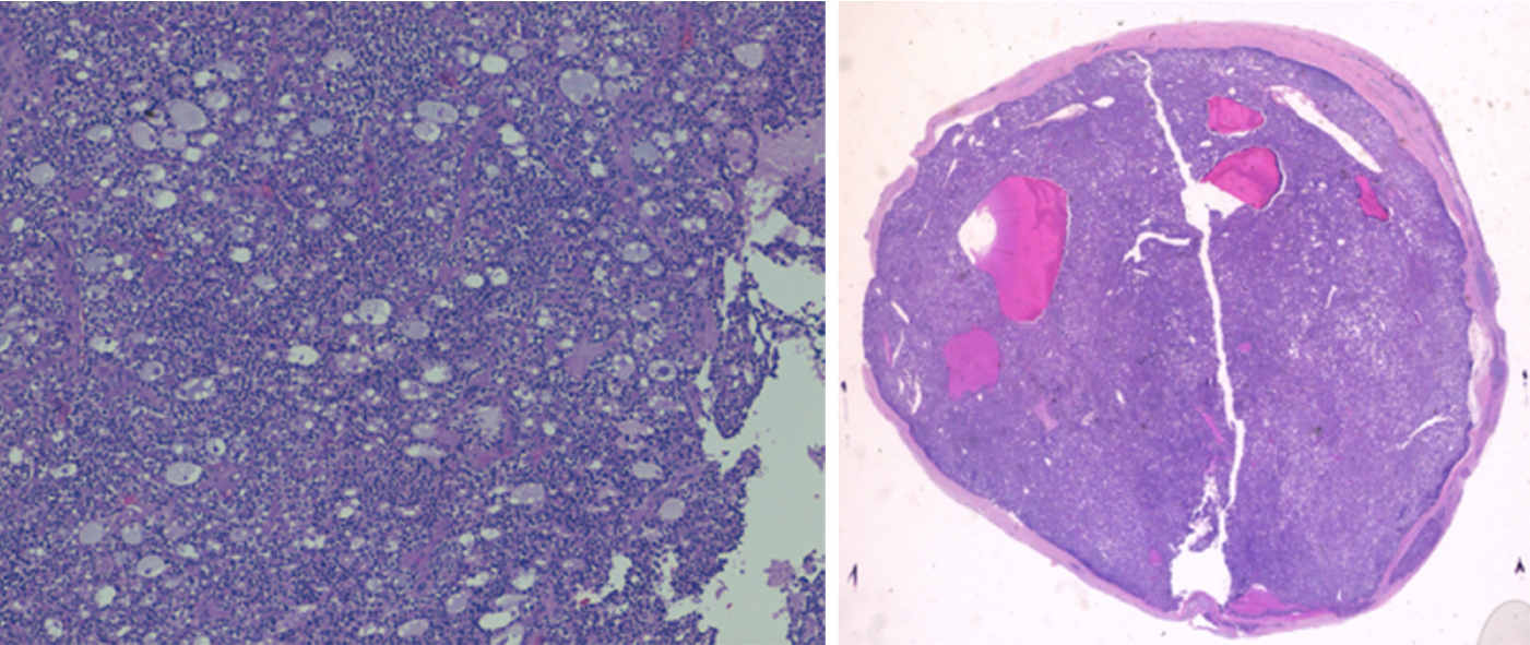
Figure 3 Microscopic findings of the parathyroid carcinoma.
It shows neoplasm organoid pattern, with moderate nuclear pleomorphism. In the last photo is seen as nests of tumor cells infiltrate the fibrous capsule.
- Citation: Triviño V, Fidalgo O, Juane A, Pombo J, Cordido F. Gonadotrophin-releasing hormone agonist-induced pituitary adenoma apoplexy and casual finding of a parathyroid carcinoma: A case report and review of literature. World J Clin Cases 2019; 7(20): 3259-3265
- URL: https://www.wjgnet.com/2307-8960/full/v7/i20/3259.htm
- DOI: https://dx.doi.org/10.12998/wjcc.v7.i20.3259


