©The Author(s) 2019.
World J Clin Cases. Jan 26, 2019; 7(2): 242-252
Published online Jan 26, 2019. doi: 10.12998/wjcc.v7.i2.242
Published online Jan 26, 2019. doi: 10.12998/wjcc.v7.i2.242
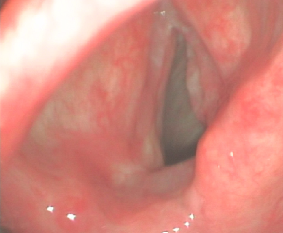
Figure 1 Laryngoscope image.
Laryngeal endoscopy shows a bulging lesion of the right true vocal cord extending from the lateral vocal cord to the anterior commissure.
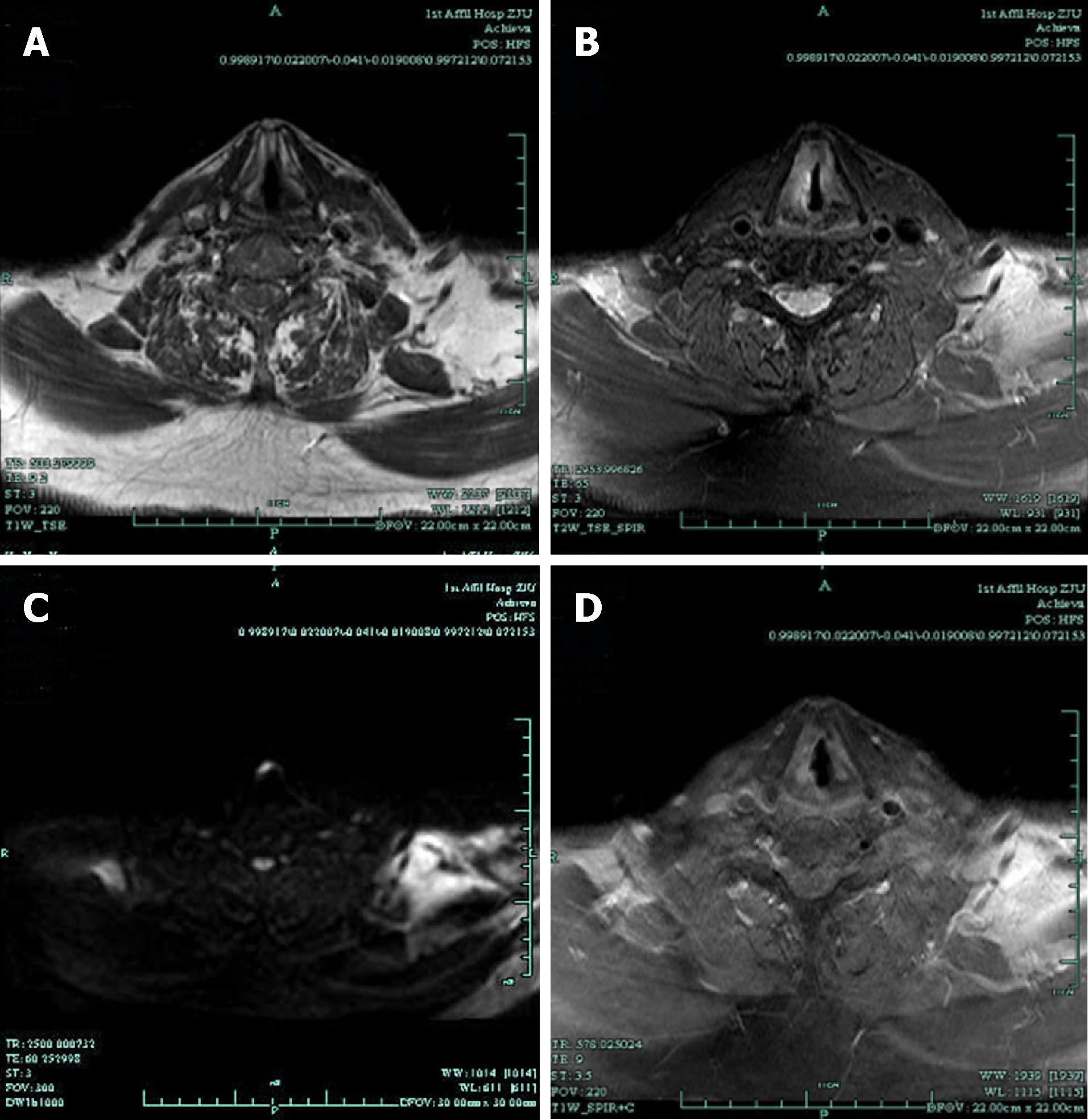
Figure 2 Magnetic resonance imaging images.
Magnetic resonance imaging (MRI) with contrast revealed that the right vocal cord was thicker than the left. A: T1-weighted imaging was isointense; B: T2-weighted imaging was hyperintense; C: Diffusion-weighted imaging was hyperintense; D: Contrast-enhanced T1-weighted MR image showed obvious enhancement.
Figure 3 Pathology images.
Pathology showed coexistence of squamous cell carcinoma (SCC) cells and neuroendocrine carcinoma (NEC) cells. A: The classic pattern of squamous epithelial dysplasia, visible keratinized beads, and infiltrative growth. Hematoxylin-eosin staining (original magnification, ×200); B: The pattern of NEC, in which most of the areas of the tumor were poorly differentiated, and diffusely infiltrated by small round cells, and the cells were heterogeneous. Hematoxylin-eosin staining (original magnification, ×200); C: The SCC component and NEC component coexist in the specimen organization. Hematoxylin-eosin staining (original magnification, ×100).
Figure 4 Immunohistochemical images.
On immunohistochemical staining (original magnification, ×200), the tumor cells were positive for (A) synaptophysin, (B) cytokeratin, (C) CD56, and (D) P63; E: The Ki67 index was up to 90%.
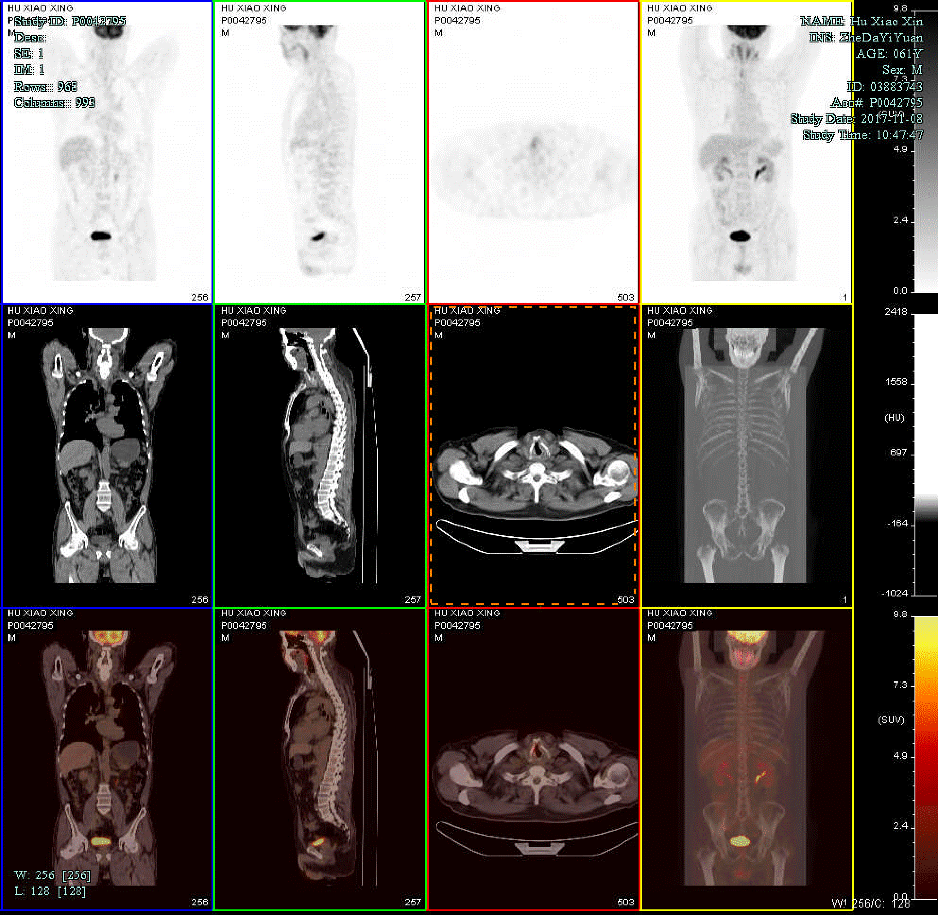
Figure 5 Postoperative positron emission tomography/computed tomography images.
Postoperative positron emission tomography/computed tomography showed high-level uptake of 18F-fluoro-2-deoxy-D-glucose (FDG) in the right vocal cord (maximum standardized uptake = 5.6), and there were no high-FDG lesions in any other part of the body.
- Citation: Yu Q, Chen YL, Zhou SH, Chen Z, Bao YY, Yang HJ, Yao HT, Ruan LX. Collision carcinoma of squamous cell carcinoma and small cell neuroendocrine carcinoma of the larynx: A case report and review of the literature. World J Clin Cases 2019; 7(2): 242-252
- URL: https://www.wjgnet.com/2307-8960/full/v7/i2/242.htm
- DOI: https://dx.doi.org/10.12998/wjcc.v7.i2.242


