©The Author(s) 2019.
World J Clin Cases. Jan 26, 2019; 7(2): 236-241
Published online Jan 26, 2019. doi: 10.12998/wjcc.v7.i2.236
Published online Jan 26, 2019. doi: 10.12998/wjcc.v7.i2.236
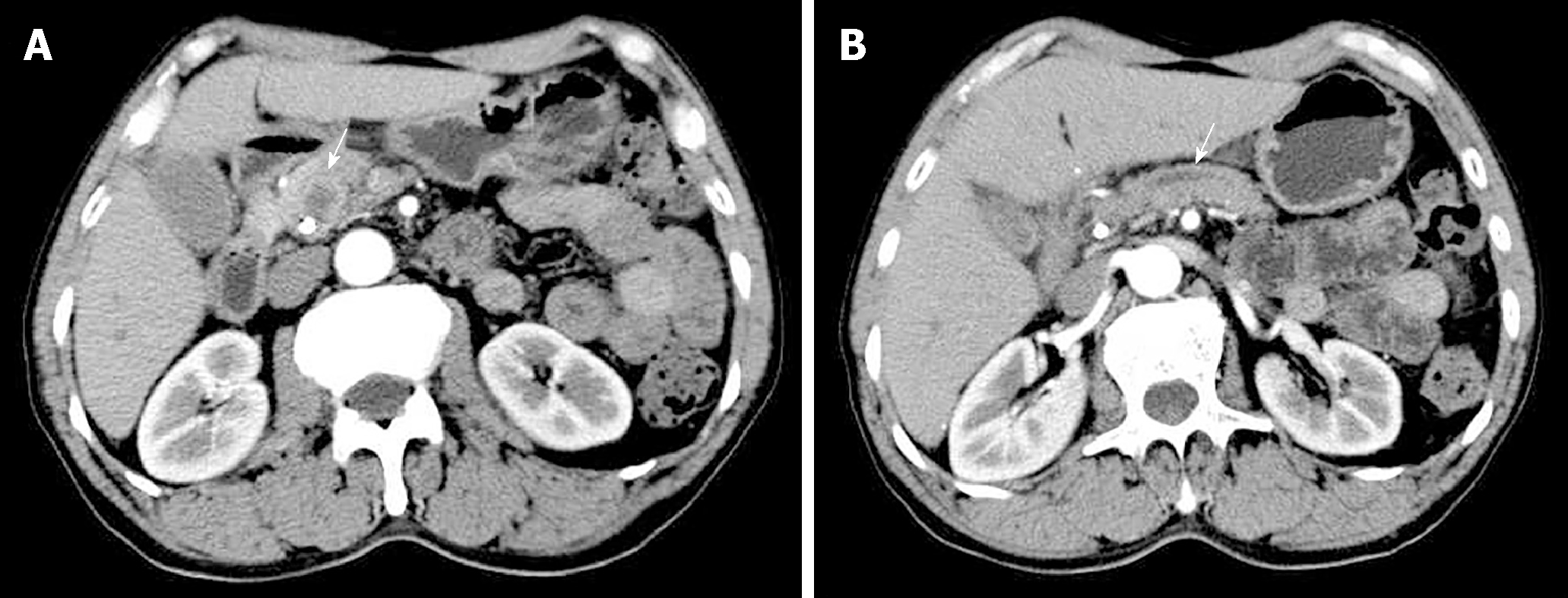
Figure 1 Arterial phase computed tomography images.
A: A low-density round mass measuring about 1.5 cm × 1.1 cm in the pancreatic head; B: Dilated pancreatic duct.
Figure 2 Pathological examination of the lesion in the pancreatic head.
A: Spindle cells with marked nuclear atypia and brisk mitotic activity arranged in a storiform or fascicular pattern (hematoxylin and eosin staining); B, C: Spindle cells exhibited strong diffuse positivity for cytokeratin 19 (B) and vimentin (C).
Figure 3 Pathological examination of the metastatic lymph nodes.
A: Spindle cells with marked nuclear atypia and brisk mitotic activity arranged in a storiform or fascicular pattern (hematoxylin and eosin staining); B, C: Spindle cells exhibited strong diffuse positivity for cytokeratin 19 (B) and vimentin (C).
- Citation: Zhou DK, Gao BQ, Zhang W, Qian XH, Ying LX, Wang WL. Sarcomatoid carcinoma of the pancreas: A case report. World J Clin Cases 2019; 7(2): 236-241
- URL: https://www.wjgnet.com/2307-8960/full/v7/i2/236.htm
- DOI: https://dx.doi.org/10.12998/wjcc.v7.i2.236


