Copyright
©The Author(s) 2019.
World J Clin Cases. Jun 6, 2019; 7(11): 1337-1343
Published online Jun 6, 2019. doi: 10.12998/wjcc.v7.i11.1337
Published online Jun 6, 2019. doi: 10.12998/wjcc.v7.i11.1337
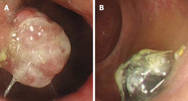
Figure 1 Preoperative colonoscopy images revealing two anorectal masses, one pedunculated mass located at the anal canal level (unpigmented, diameter 2.
5 cm) (A), and the other sessile mass 3 cm above the dentate line (pigmented, diameter 2 cm) (B).
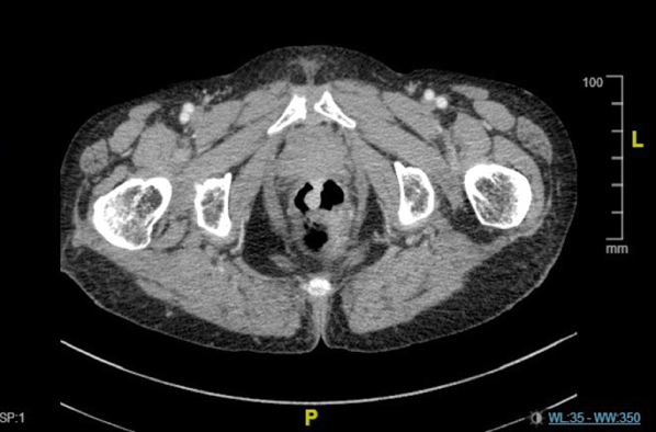
Figure 2 Enhanced pelvic computed tomography image revealing a pedunculated mass from the anterior wall of the rectum, and the other sessile mass from the side wall of the rectum.
Both masses invaded into muscular layer and were enhanced at the arterial phase.
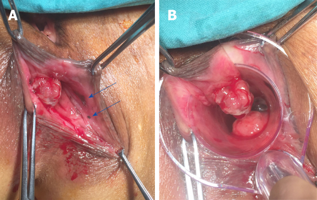
Figure 3 Transanal exploration of anorectal masses.
A: Derived from the anterior wall of the rectum, one pedunculated mass appeared at the anal canal level without melanin pigmentation. Two mucosal melanic zones were found at the anal canal level (blue arrows); B: Another pigmented mass was 3 cm above the dentate line under anoscope vision.
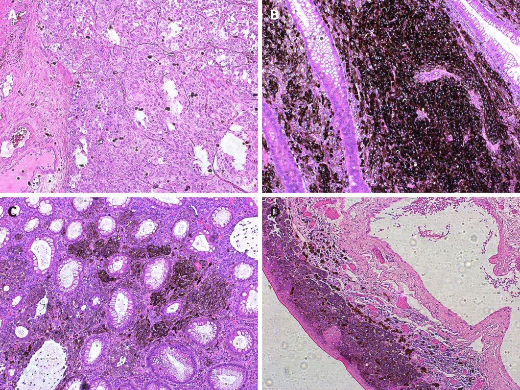
Figure 4 Histologic illustration of the four lesions in this case (HE staining, 10×).
A: Pedunculated mass located at the anal canal level. Although this lesion appeared unpigmented via visualization, scattered round atypical pigmented cells were found microscopically; B: The sessile mass 3 cm above the dentate line showed densely distributed pigmented cells; C and D: Two mucosal melanic zones were analyzed. Infiltration of atypical pigmented cells was found distributed in the mucosal and submucosal layers.
- Citation: Cai YT, Cao LC, Zhu CF, Zhao F, Tian BX, Guo SY. Multiple synchronous anorectal melanomas with different colors: A case report. World J Clin Cases 2019; 7(11): 1337-1343
- URL: https://www.wjgnet.com/2307-8960/full/v7/i11/1337.htm
- DOI: https://dx.doi.org/10.12998/wjcc.v7.i11.1337


