©The Author(s) 2019.
World J Clin Cases. Jan 6, 2019; 7(1): 19-27
Published online Jan 6, 2019. doi: 10.12998/wjcc.v7.i1.19
Published online Jan 6, 2019. doi: 10.12998/wjcc.v7.i1.19
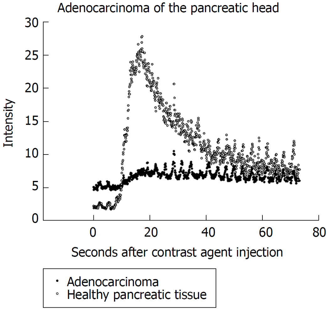
Figure 1 During contrast enhanced harmonic endoscopic ultrasound procedures patients received 5 mL SonoVue® contrast agent.
The figure shows an example of ultrasound signal intensity curves of healthy pancreatic tissue and pancreatic adenocarcinoma.
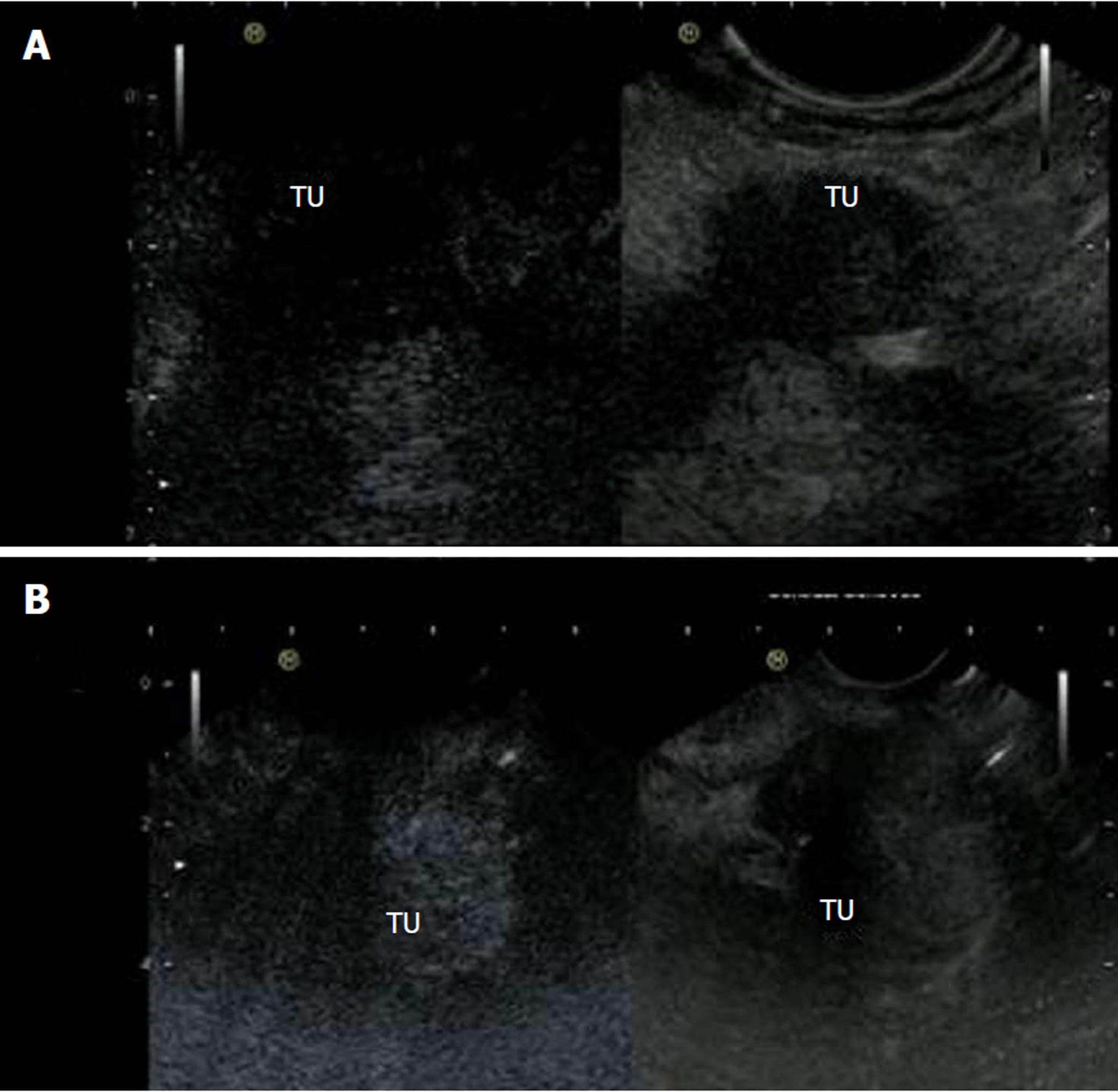
Figure 2 Contrast enhanced harmonic endoscopic ultrasound images of hypoenhanced pancreatic adenocarcinoma and hyperenhancement of neuroendocrine carcinoma.
A: Contrast enhanced harmonic endoscopic ultrasound (CEH-EUS) images of hypoenhanced pancreatic adenocarcinoma; B: CEH-EUS images of neuroendocrine carcinoma. TU: Tumor left side.
- Citation: Kannengiesser K, Mahlke R, Petersen F, Peters A, Kucharzik T, Maaser C. Instant evaluation of contrast enhanced endoscopic ultrasound helps to differentiate various solid pancreatic lesions in daily routine. World J Clin Cases 2019; 7(1): 19-27
- URL: https://www.wjgnet.com/2307-8960/full/v7/i1/19.htm
- DOI: https://dx.doi.org/10.12998/wjcc.v7.i1.19


