©The Author(s) 2018.
World J Clin Cases. Jul 16, 2018; 6(7): 156-160
Published online Jul 16, 2018. doi: 10.12998/wjcc.v6.i7.156
Published online Jul 16, 2018. doi: 10.12998/wjcc.v6.i7.156
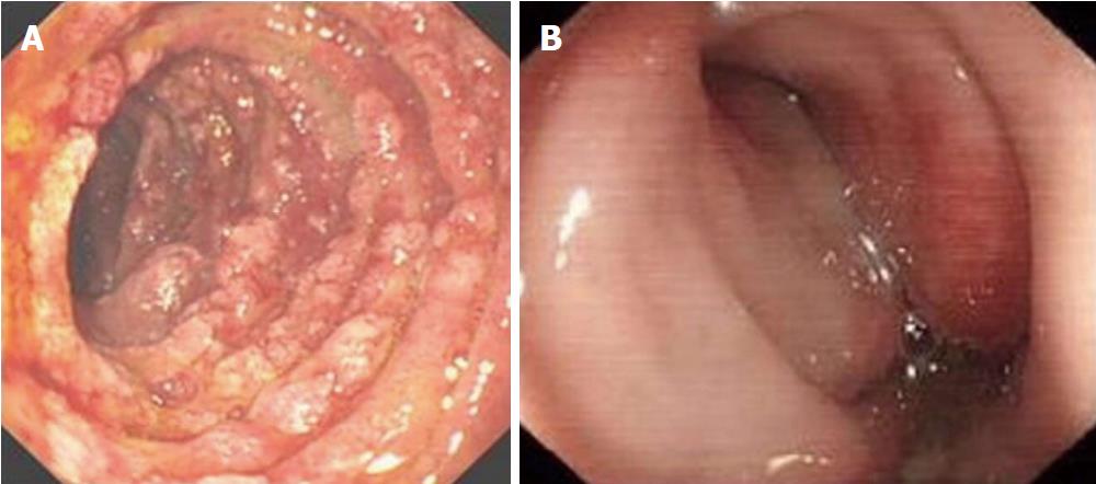
Figure 1 Endoscopic examination.
A: Endoscopic view of the duodenal mucosa shows erosions, exudates, and mucosal rings; B: Endoscopic examination revealed erosions, hyperemia, and swelling of the rectal mucosal.
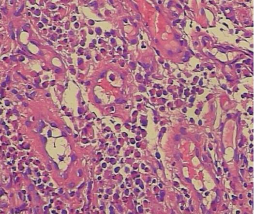
Figure 2 Histological examination demonstrates histological findings of eosinophilic infiltration of the duodenal mucosa (HE × 200).
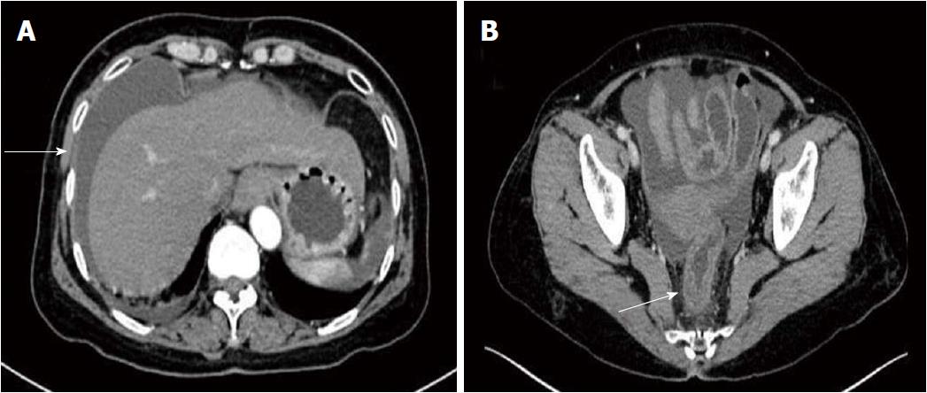
Figure 3 Radiological images of the abdomen.
A: Computed tomography (CT) of the abdomen shows massive ascites (arrow); B: Abdominal CT shows accompanied local mild thickening of the right rear rectum wall (arrow).
- Citation: Shi L, Jia QH, Liu FJ, Guan H, Jiang ZY. Massive hemorrhagic ascites: A rare presentation of eosinophilic gastroenteritis. World J Clin Cases 2018; 6(7): 156-160
- URL: https://www.wjgnet.com/2307-8960/full/v6/i7/156.htm
- DOI: https://dx.doi.org/10.12998/wjcc.v6.i7.156


