©The Author(s) 2018.
World J Clin Cases. Jul 16, 2018; 6(7): 143-149
Published online Jul 16, 2018. doi: 10.12998/wjcc.v6.i7.143
Published online Jul 16, 2018. doi: 10.12998/wjcc.v6.i7.143
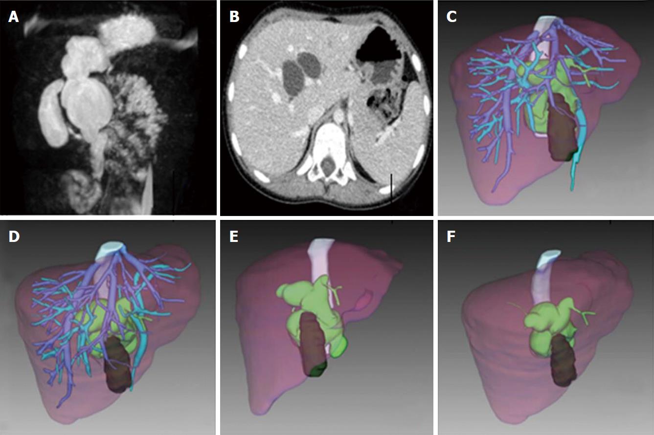
Figure 1 Preoperation images.
Magnetic resonance cholangiopancreatography, euglycemic hyperinsulinemic clamp technique and 3D reconstruction. A: Preoperation MRCP shows type IVa CCs; B: Preoperation EHCT shows CCs involves intrahepatic bile duct; C: Liver and CCs 3D view from front; D: Liver and CCs 3D view from middle hepatic vein (MHV); E: Liver and CCs 3D view from front without vessels; F: Liver and CCs 3D view from middle hepatic vein (MHV) without vessels. CCS: Congenital choledochal cysts.
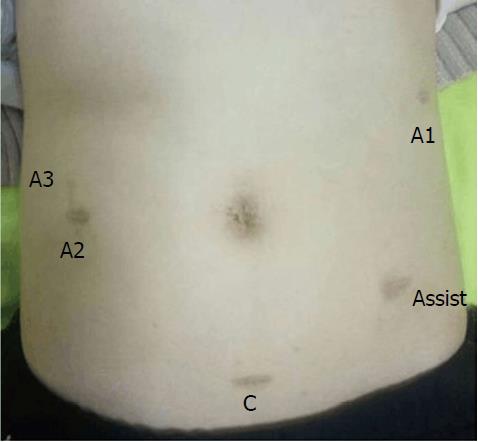
Figure 2 Postoperation images.
C: Camera port; A1/2/3: Arm 1/2/3 port; Assist: Assistant/accessory port.
- Citation: Wang XQ, Xu SJ, Wang Z, Xiao YH, Xu J, Wang ZD, Chen DX. Robotic-assisted surgery for pediatric choledochal cyst: Case report and literature review. World J Clin Cases 2018; 6(7): 143-149
- URL: https://www.wjgnet.com/2307-8960/full/v6/i7/143.htm
- DOI: https://dx.doi.org/10.12998/wjcc.v6.i7.143


