©The Author(s) 2018.
World J Clin Cases. Jun 16, 2018; 6(6): 132-138
Published online Jun 16, 2018. doi: 10.12998/wjcc.v6.i6.132
Published online Jun 16, 2018. doi: 10.12998/wjcc.v6.i6.132
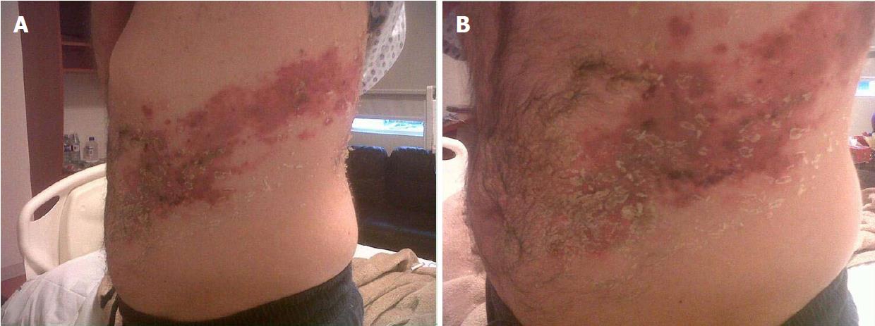
Figure 1 Distended abdomen with vesicular eruption involving the left T7-T10 dermatomes (A and B).
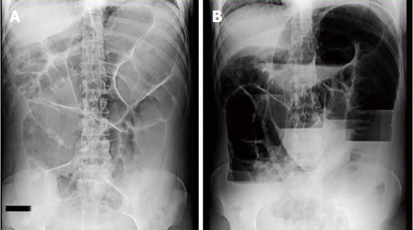
Figure 2 Plain abdominal radiograph revealing defuses dilatation of the small bowel and air-fluid levels.
A: Supine view; B: Upright view.
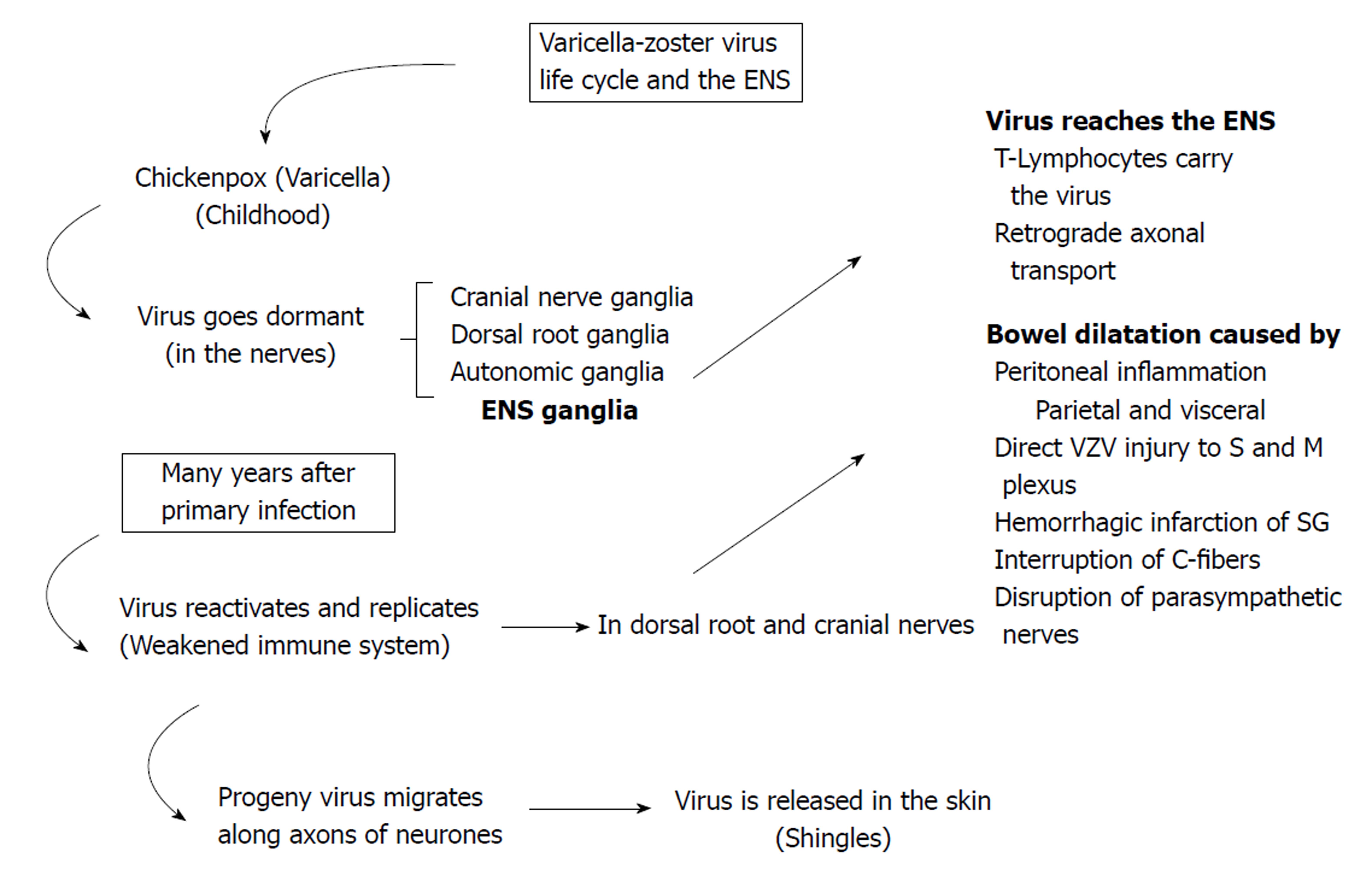
Figure 3 The varicella-zoster virus life cycle.
VZV first contact usually takes place early in childhood. Then, the virus goes dormant in the nerves. Years after, endogenous virus reactivation occurs in the ganglion. Virus replicates and migrates to the skin or the ENS, causing either shingles or paralytic ileus, respectively. ENS: Enteric nervous system; S and M: Submucosal and myenteric; SG: Sympathetic ganglia; VZV: Varicella-zoster virus.
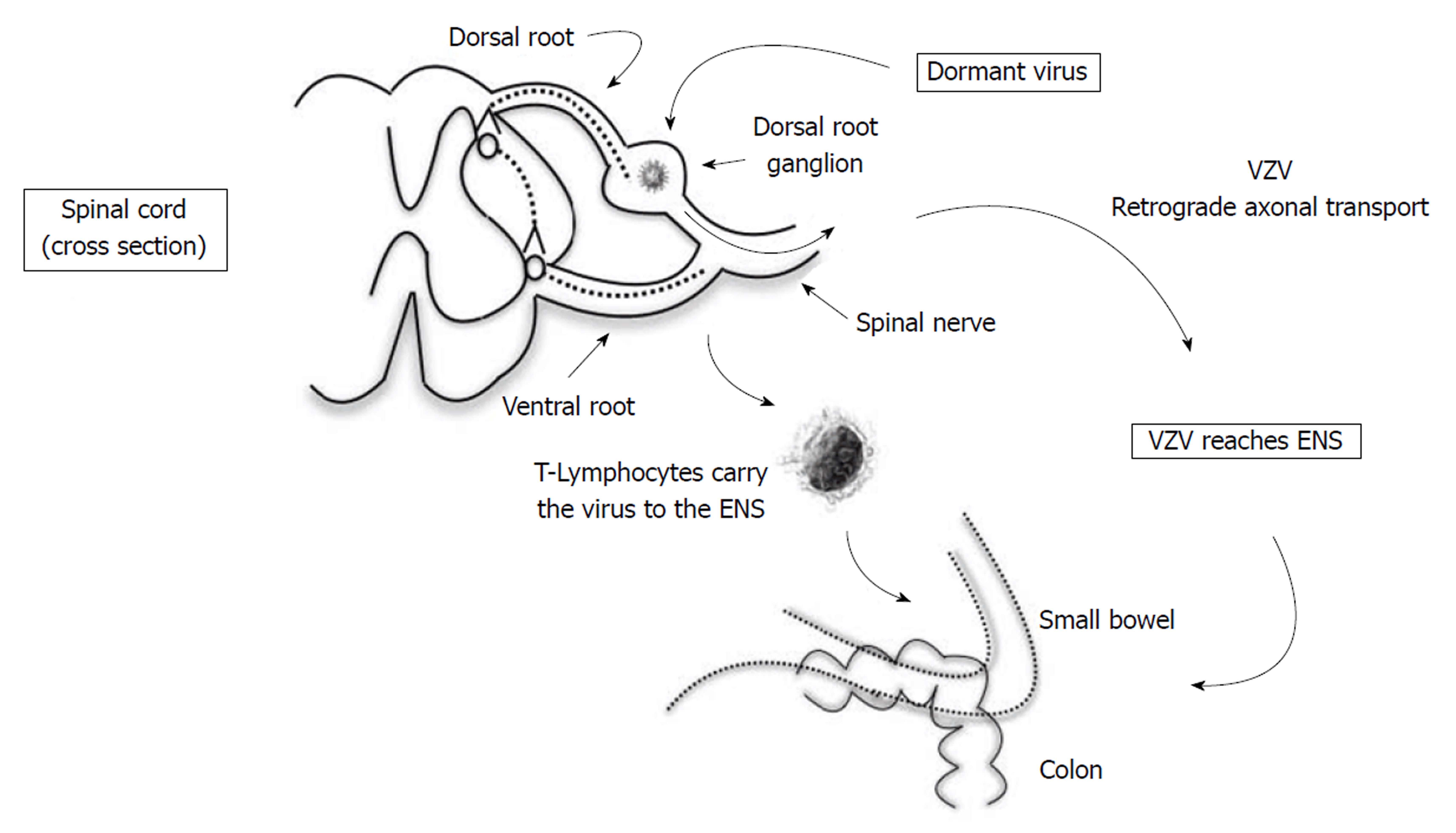
Figure 4 Possible theory explains how varicella-zoster virus reaches the enteric nervous system, causing direct injury to submucosal and myenteric plexus.
Either T-Lymphocytes carry the virus from dorsal root ganglion or progeny virus migrates along the axons of neurones in a retrograde fashion. ENS: Enteric nervous system; VZV: Varicella-zoster virus.
- Citation: Anaya-Prado R, Pérez-Navarro JV, Corona-Nakamura A, Anaya-Fernández MM, Anaya-Fernández R, Izaguirre-Pérez ME. Intestinal pseudo-obstruction caused by herpes zoster: Case report and pathophysiology. World J Clin Cases 2018; 6(6): 132-138
- URL: https://www.wjgnet.com/2307-8960/full/v6/i6/132.htm
- DOI: https://dx.doi.org/10.12998/wjcc.v6.i6.132


