Copyright
©The Author(s) 2018.
World J Clin Cases. Dec 6, 2018; 6(15): 1036-1041
Published online Dec 6, 2018. doi: 10.12998/wjcc.v6.i15.1036
Published online Dec 6, 2018. doi: 10.12998/wjcc.v6.i15.1036
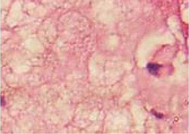
Figure 1 Results of pathological examination.
Subcutaneous nodules, fat necrosis (× 200)
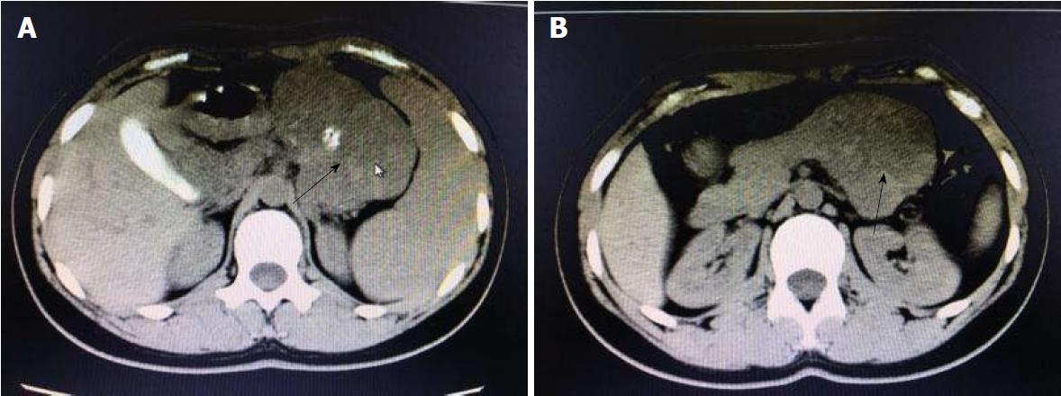
Figure 2 Findings of plain abdominal computed tomography scan.
A: Plain abdominal computed tomography reveals a soft-tissue mass in the body and tail of pancreas; B: It has a well-defined border and sized 8.5 cm × 6.1 cm × 6.5 cm, with heterogeneous signal intensity patches and nodular calcifications. It is closely associated with gastric wall. Arrow indicates the mass.
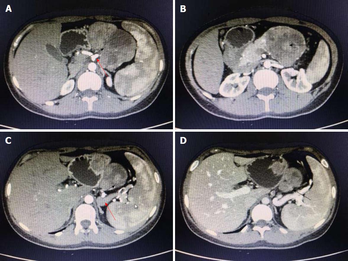
Figure 3 Findings of contrast-enhanced abdominal computed tomography scan.
A and B: Contrast-enhanced abdominal computed tomography reveals a mixed density mass with solid and cystic components, sized 6.8 cm × 7.8 cm × 8.8 cm, in the body and tail of the pancreas, with a well-defined border. Nodular calcifications are found inside the lesion; C: The solid component is obviously enhanced, although the degree of enhancement is lower than the normal pancreatic parenchyma; D: The spleen is slightly larger, and the splenic vein is compressed. Multiple collateral circulations are seen in the left upper abdomen. The lesion is fed by the common hepatic artery and a branch of splenic artery (red arrows in A and C, black arrows indicate the mass).
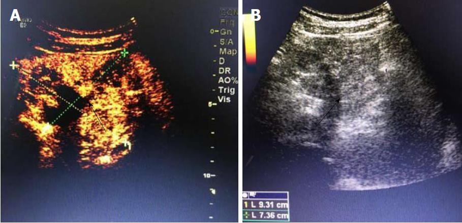
Figure 4 Findings of abdominal contrast-enhanced ultrasonography.
A: Abdominal contrast-enhanced ultrasonography shows an abnormal morphology of the pancreas. A mass with inhomogeneous echo, sized about 9.3 cm × 7.2 cm, is found in the anterior pancreatic body; it has a well-defined border and regular shape, but the border with the parenchyma of pancreatic body and tail is poorly defined, with flocculent echo and strong spotty or patchy echo in the central part; B: Color Doppler flow imaging showed a few low-speed arteriovenous blood flow signals, whereas the echoes are homogeneous in the remaining parenchyma. The main pancreatic duct is not dilated. Black arrows indicate the mass.
Figure 5 Specimen of the resected pancreas and results of pathological examination.
A: Specimen of the resected pancreas; B and C: A solid pseudopapillary tumor of the pancreas, sized about 8.2 cm × 8 cm × 5 cm, showed infiltrative growth and necrosis. No vascular or neural invasion was visible. Immunophenotyping showed synaptophysin(+), CD56(+), CD10(+), β-catenin(+), vimentin(+), AE1/AE3(+), PR(+), plasma chromogranin A(-), E-cadherin(-), and Ki-67(+/3%). B: Hematoxylin and eosin staining (× 100); C: Immunohistochemistry (× 100).
- Citation: Zhang MY, Tian BL. Pancreatic panniculitis and solid pseudopapillary tumor of the pancreas: A case report. World J Clin Cases 2018; 6(15): 1036-1041
- URL: https://www.wjgnet.com/2307-8960/full/v6/i15/1036.htm
- DOI: https://dx.doi.org/10.12998/wjcc.v6.i15.1036


