Copyright
©The Author(s) 2018.
World J Clin Cases. Sep 26, 2018; 6(10): 365-372
Published online Sep 26, 2018. doi: 10.12998/wjcc.v6.i10.365
Published online Sep 26, 2018. doi: 10.12998/wjcc.v6.i10.365
Figure 1 Mini open incision method.
A: Local anesthetic application to the incision line; B: The standard incision starts from the distal volar wrinkle, passes between the thenar and hypothenar region 2-3 mm medially to thenar wrinkle and extends 2-3 cm distally to the lateral side of the third finger; C: Placement of the skin retractor after sharp dissection.
Figure 2 Extended mini-open incision technique in a patient previously operated on using the uniprotal endoscopic method.
A: Endoscopic portal scar over the distal wrist wrinkle (red arrow); B: Incomplete incision of the transverse carpal ligament and compression on the median nerve (black arrow); C: The incision is completed and the median nerve is fully decompressed.
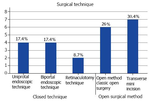
Figure 3 Ten (43.
4%) recurrent cases were previously operated with closed technique (uniprotal endoscopic technique in four, biportal endoscopic technique in four and retinaculotomy technique in two cases). Six (26%) recurrent cases were previously operated with open surgical method and seven (30.4%) recurrent cases were previously operated with transverse mini incision.
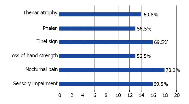
Figure 4 Thenar atrophy in 14 (60.
8%) cases. Phalen test was positive in 13 (56.5%) cases. Tinel sign was found in 16 (69.5%) cases. Loss of hand strength in 13 (56.5%) cases, nocturnal pain in 18 (78.2%) cases and sensory impairment was detected in 16 (69.5%) cases.
- Citation: Eroğlu A, Sarı E, Topuz AK, Şimşek H, Pusat S. Recurrent carpal tunnel syndrome: Evaluation and treatment of the possible causes. World J Clin Cases 2018; 6(10): 365-372
- URL: https://www.wjgnet.com/2307-8960/full/v6/i10/365.htm
- DOI: https://dx.doi.org/10.12998/wjcc.v6.i10.365


