©The Author(s) 2017.
World J Clin Cases. Jun 16, 2017; 5(6): 247-253
Published online Jun 16, 2017. doi: 10.12998/wjcc.v5.i6.247
Published online Jun 16, 2017. doi: 10.12998/wjcc.v5.i6.247
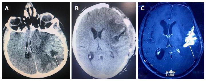
Figure 1 Showing radiological scans and findings.
A: Computed tomography (CT) scan at initial diagnosis suggestive of glioma; B: Post-operative image of CT scan with adequate tumor excision and reduced mass effect; C: Magnetic resonance image after 18 mo showing the progressive recurrence of tumor.
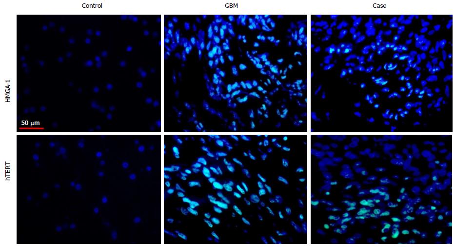
Figure 2 Immunofluorescence based immuno-histochemistry analysis and interpretation for human telomerase reverse transcriptase and high mobility group-A1: Control sample showed negative expression of both markers; intense immune-reactivity of high mobility group-A1 and human telomerase reverse transcriptase in Glioblastoma-multiforme and moderate expression in our case.
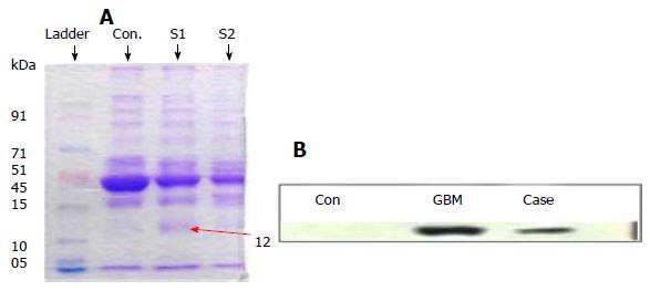
Figure 3 High mobility group-A1.
A: SDS-PAGE protein profile of serum samples; B: Western blot expression for high mobility group-A1 antibody in control serum sample (Con), GBM and present case. GBM: Glioblastoma-multiforme.
- Citation: Gandhi P, Khare R, Garg N, Sorte S. Immunophenotypic signature of primary glioblastoma multiforme: A case of extended progression free survival. World J Clin Cases 2017; 5(6): 247-253
- URL: https://www.wjgnet.com/2307-8960/full/v5/i6/247.htm
- DOI: https://dx.doi.org/10.12998/wjcc.v5.i6.247


