©The Author(s) 2017.
World J Clin Cases. Apr 16, 2017; 5(4): 148-152
Published online Apr 16, 2017. doi: 10.12998/wjcc.v5.i4.148
Published online Apr 16, 2017. doi: 10.12998/wjcc.v5.i4.148
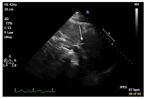
Figure 1 Echocardiography - modified subcostal view showing liner foreign body in the right atrium.
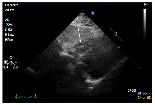
Figure 2 Echocardiography subcostal view showing liner foreign body in the right ventricle.
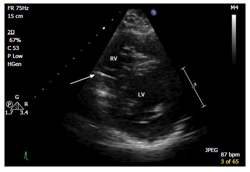
Figure 3 Echocardiography short axis view showing liner foreign body in the right ventricle.
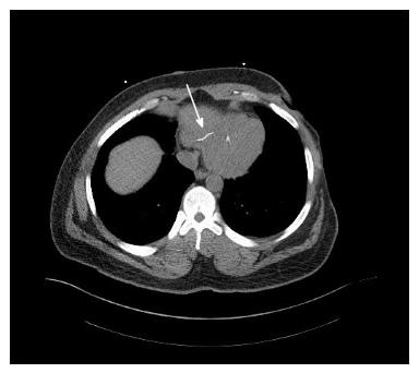
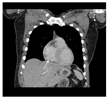
Figure 4 Computed tomography of the chest showing liner foreign body in the right ventricle.
Figure 5 Computed tomography of the chest showing liner foreign body in the right ventricle.
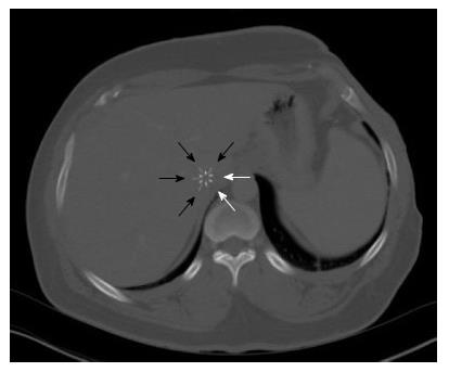
Figure 6 Computed tomography of the abdomen showing missing limbs of the filter.
Black arrows showing 4 outer limbs and white arrows showing the position of missing outer limbs.
- Citation: Sivasambu B, Kabirdas D, Movahed A. Incidental echocardiographic finding: Fractured inferior vena cava filter. World J Clin Cases 2017; 5(4): 148-152
- URL: https://www.wjgnet.com/2307-8960/full/v5/i4/148.htm
- DOI: https://dx.doi.org/10.12998/wjcc.v5.i4.148


