©The Author(s) 2016.
World J Clin Cases. Mar 16, 2016; 4(3): 94-98
Published online Mar 16, 2016. doi: 10.12998/wjcc.v4.i3.94
Published online Mar 16, 2016. doi: 10.12998/wjcc.v4.i3.94
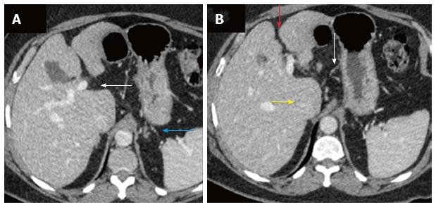
Figure 1 Triple phase dynamic contrast enhanced computed tomography performed for the upper abdomen showed features of chronic liver disease, predominantly on the portal venous phase.
A: Subtle sign of periportal space widening (white arrow) and few small collaterals in the peri-gastric location (blue arrow) suggesting features of chronic liver disease on computed tomography scan; B: Irregular liver margins seen better in the inter-lobar fissure (red arrow) with mild caudate enlargement (yellow arrow) and perigastric collaterals (white arrow).
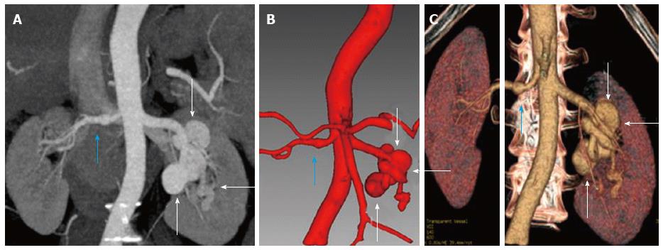
Figure 2 Reformatted images of the bilateral renal artery aneurysms, in right main renal artery and the segmental left renal artery divisions.
A: Coronal minimum intensity projection reformat of arterial phase of the triple phase computed tomography (CT) scan showing right main renal artery aneurysm (blue arrow) and multiple left segmental division branch aneurysms (white arrows); B: Coronal reformats of arterial phase of the CT angiogram performed using Myrian Intrasense software for liver showing right main renal artery aneurysm (blue arrow) and multiple left segmental division branch aneurysms (white arrows); C: Coronal reformats of arterial phase of the CT angiogram performed using Myrian Intrasense software for liver showing right main renal artery aneurysm (blue arrow) and multiple left segmental division branch aneurysms (white arrows).
- Citation: Laroia ST, Lata S. Hypertension in the liver clinic - polyarteritis nodosa in a patient with hepatitis B. World J Clin Cases 2016; 4(3): 94-98
- URL: https://www.wjgnet.com/2307-8960/full/v4/i3/94.htm
- DOI: https://dx.doi.org/10.12998/wjcc.v4.i3.94


