Copyright
©The Author(s) 2015.
World J Clin Cases. Sep 16, 2015; 3(9): 843-847
Published online Sep 16, 2015. doi: 10.12998/wjcc.v3.i9.843
Published online Sep 16, 2015. doi: 10.12998/wjcc.v3.i9.843
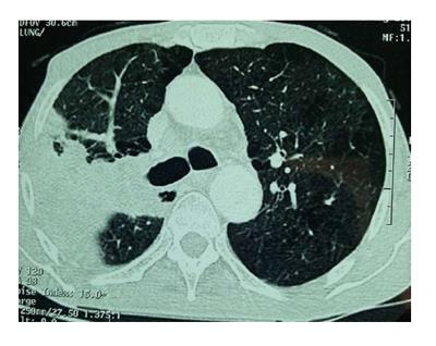
Figure 1 Thoracic computed tomography scan showed a mass in the upper lobe of the right lung with invasion of the mediastinal pleura and ipsilateral mediastinal lymphadenopathy.
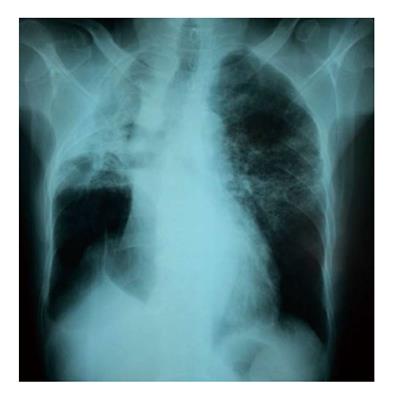
Figure 2 Chest X-ray performed at the second patient’s admission showing the right upper lobe lung tumor, and nodular parenchymal infiltrates in the left upper lobe lung with a small right pleural effusion.
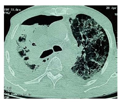
Figure 3 Thoracic computed tomography scan showing a primitive tumor with widespread thin-walled cysts and nodules throughout the lungs but most prominent at the left lung.
There were also a small right apical pneumothorax and a small bilateral pleural effusion.
-
Citation: Msaad S, Yangui I, Bahloul N, Abid N, Koubaa M, Hentati Y, Ben Jemaa M, Kammoun S. Do inhaled corticosteroids increase the risk of
Pneumocystis pneumonia in people with lung cancer? World J Clin Cases 2015; 3(9): 843-847 - URL: https://www.wjgnet.com/2307-8960/full/v3/i9/843.htm
- DOI: https://dx.doi.org/10.12998/wjcc.v3.i9.843


