Copyright
©The Author(s) 2015.
World J Clin Cases. Aug 16, 2015; 3(8): 751-756
Published online Aug 16, 2015. doi: 10.12998/wjcc.v3.i8.751
Published online Aug 16, 2015. doi: 10.12998/wjcc.v3.i8.751
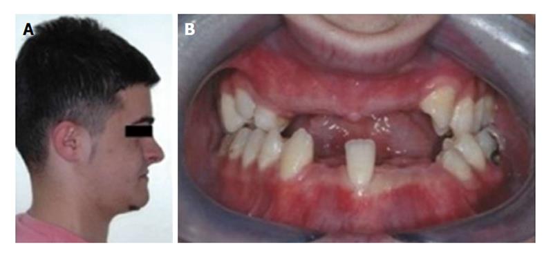
Figure 1 Clinical appearance before orthodontic treatment.
A: Profile photo of the patient before orthodontic treatment; B: There were several missing teeth in the upper and lower dental arches.
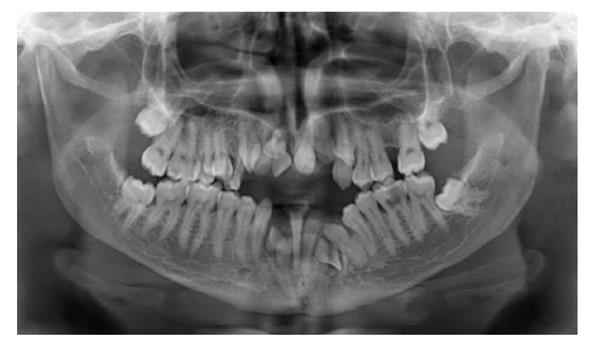
Figure 2 Pre-orthodontic panoromic radiography reveals multiple impacted permanent teeth with impacted supernumerary teeth and retained deciduous teeth.
Ten permanent teeth were impacted. There were 2 supernumerary impacted teeth in the mandible and 1 supernumerary impacted tooth in the maxilla.
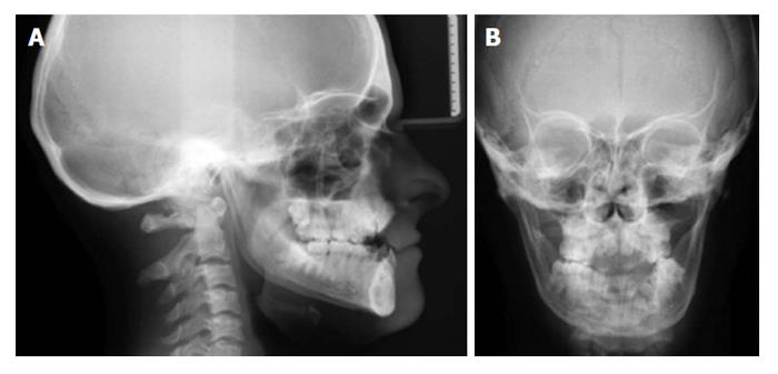
Figure 3 Sephalometric (A) and posterio-anterior radiographies (B) of the patient before orthodontic treatment.
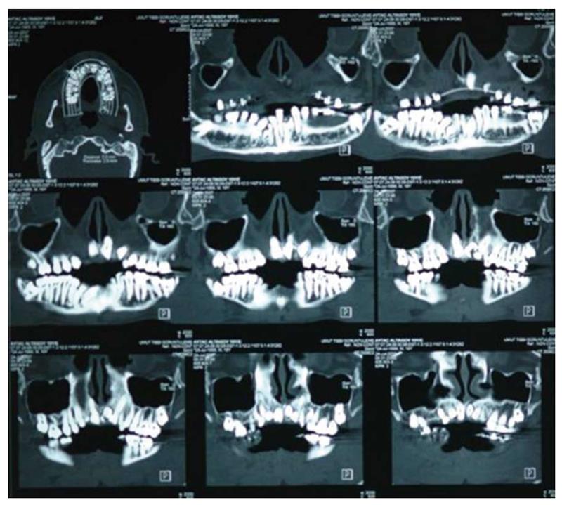
Figure 4 Frontal cone beam computerized tomography sections show impacted permanent and supernumerary teeth in detail.
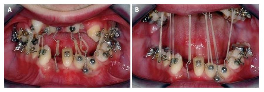
Figure 5 Intraoral elastics were applied between impacted teeth.
A: Elastics remained passive when mouth is closed; B: They were activated when patient opened his mouth for daily activity such as speaking or drinking.
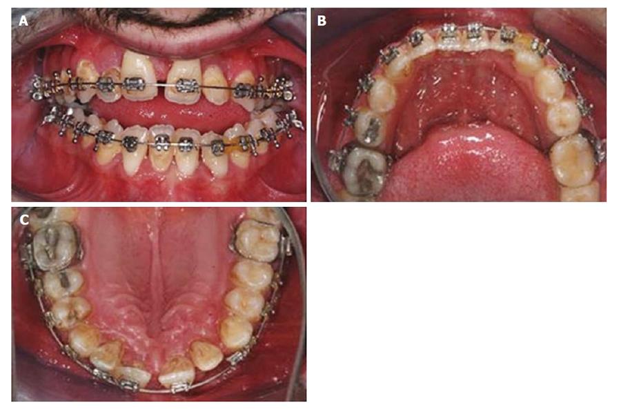
Figure 6 Clinical intraoral appearance after orthodontic treatment.
A: The intraoral front-view of upper and lower dental archs after orthodontic traction and leveling treatment; B: Occlusal view of lower dental arch; C: Occlusal view of upper dental arch.
Figure 7 Radiographic and clinical evaluation after orthodontic treatment.
A: Panaromic radiography after orthodontic leveling phase; B: Sephalometric radiography shows anterior open-bite malocclusion; C: Profile photograph of the patient after orthodontic leveling phase.
Figure 8 Radiographic and clinical evaluation after 5 years follow-up period.
A: Panaromic radiography 5 years after orthognathic surgery reveals preserved and unchanged dental alignment; B: Dental class I occlusal relationship is clearly seen on sephalometric radiography; C: Profile appearance of the patient seems satisfying.
- Citation: Çimen E, Dereci &, Tüzüner-Öncül AM, Yazıcıoğlu D, Özdiler E, Şenol A, Sayan NB. Combined surgical-orthodontic rehabilitation of cleidocranial dysplasia: 5 years follow-up. World J Clin Cases 2015; 3(8): 751-756
- URL: https://www.wjgnet.com/2307-8960/full/v3/i8/751.htm
- DOI: https://dx.doi.org/10.12998/wjcc.v3.i8.751


