©The Author(s) 2015.
World J Clin Cases. Jun 16, 2015; 3(6): 514-518
Published online Jun 16, 2015. doi: 10.12998/wjcc.v3.i6.514
Published online Jun 16, 2015. doi: 10.12998/wjcc.v3.i6.514
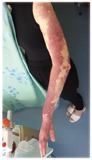
Figure 1 Vascular malformation, left upper extremity of mother.
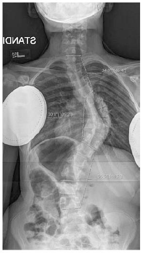
Figure 2 Pre-operative AP X-ray.
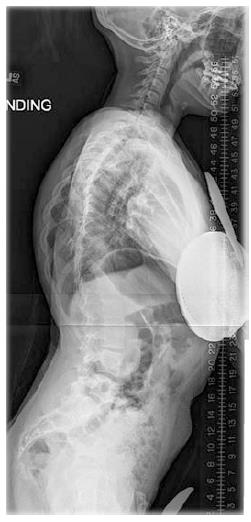
Figure 3 Pre-operative lateral X-ray.
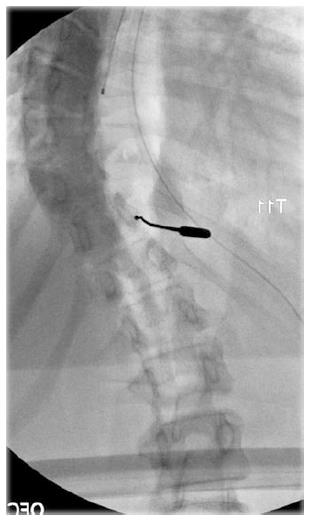
Figure 4 Intraoperative X-ray from initial procedure at right T11 pedicle.
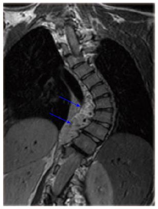
Figure 5 Coronal T2-weighted magnetic resonance imaging.
Figure 6 Axial T2-weighted magnetic resonance imaging.
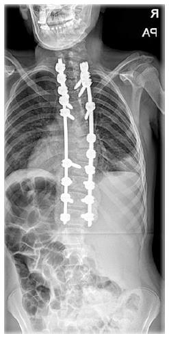
Figure 7 Post-operative AP X-ray.
- Citation: Swarup I, Bjerke-Kroll BT, Cunningham ME. Paraspinous hemolymphangioma associated with adolescent scoliosis. World J Clin Cases 2015; 3(6): 514-518
- URL: https://www.wjgnet.com/2307-8960/full/v3/i6/514.htm
- DOI: https://dx.doi.org/10.12998/wjcc.v3.i6.514


